3B2Q

Intermediate position of ATP on its trail to the binding pocket inside the subunit B mutant R416W of the energy converter A1Ao ATP synthase


Experimental Data Snapshot

  • Method: X-RAY DIFFRACTION
  • Resolution: 2.10 Å
  • R-Value Free: 0.237 
  • R-Value Work: 0.189 
  • R-Value Observed: 0.189 

wwPDB Validation   3D Report Full Report


Ligand Structure Quality Assessment 


This is version 1.4 of the entry. See complete history


Literature

Spectroscopic and crystallographic studies of the mutant R416W give insight into the nucleotide binding traits of subunit B of the A1Ao ATP synthase

Kumar, A.Manimekalai, M.S.S.Balakrishna, A.M.Hunke, C.Weigelt, S.Sewald, N.Gruber, G.

(2009) Proteins 75: 807-819

  • DOI: https://doi.org/10.1002/prot.22289
  • Primary Citation of Related Structures:  
    2RKW, 3B2Q

  • PubMed Abstract: 

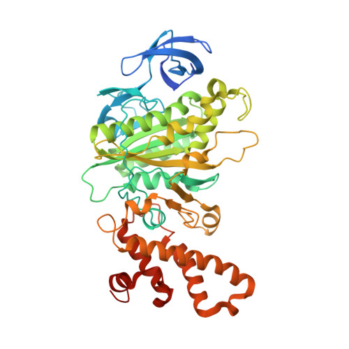
    A strategically placed tryptophan in position of Arg416 was used as an optical probe to monitor adenosine triphosphate and adenosine-diphosphate binding to subunit B of the A(1)A(O) adenosine triphosphate (ATP) synthase from Methanosarcina mazei Gö1. Tryptophan fluorescence and fluorescence correlation spectroscopy gave binding constants indicating a preferred binding of ATP over ADP to the protein. The X-ray crystal structure of the R416W mutant protein in the presence of ATP was solved to 2.1 A resolution, showing the substituted Trp-residue inside the predicted adenine-binding pocket. The cocrystallized ATP molecule could be trapped in a so-called transition nucleotide-binding state. The high resolution structure shows the phosphate residues of the ATP near the P-loop region (S150-E158) and its adenine ring forms pi-pi interaction with Phe149. This transition binding position of ATP could be confirmed by tryptophan emission spectra using the subunit B mutant F149W. The trapped ATP position, similar to the one of the binding region of the antibiotic efrapeptin in F(1)F(O) ATP synthases, is discussed in light of a transition nucleotide-binding state of ATP while on its way to the final binding pocket. Finally, the inhibitory effect of efrapeptin C in ATPase activity of a reconstituted A(3)B(3)- and A(3)B(R416W)(3)-subcomplex, composed of subunit A and the B subunit mutant R416W, of the A(1)A(O) ATP synthase is shown.


  • Organizational Affiliation

    Division of Structural and Computational Biology, Nanyang Technological University, School of Biological Sciences, 60 Nanyang Drive, Singapore 637551, Republic of Singapore.


Macromolecules
Find similar proteins by:  (by identity cutoff)  |  3D Structure
Entity ID: 1
MoleculeChains Sequence LengthOrganismDetailsImage
V-type ATP synthase beta chain
A, B
469Methanosarcina mazeiMutation(s): 1 
Gene Names: atpB
EC: 3.6.3.14
UniProt
Find proteins for Q60187 (Methanosarcina mazei (strain ATCC BAA-159 / DSM 3647 / Goe1 / Go1 / JCM 11833 / OCM 88))
Explore Q60187 
Go to UniProtKB:  Q60187
Entity Groups  
Sequence Clusters30% Identity50% Identity70% Identity90% Identity95% Identity100% Identity
UniProt GroupQ60187
Sequence Annotations
Expand
  • Reference Sequence
Small Molecules
Ligands 3 Unique
IDChains Name / Formula / InChI Key2D Diagram3D Interactions
ATP
Query on ATP

Download Ideal Coordinates CCD File 
C [auth A]ADENOSINE-5'-TRIPHOSPHATE
C10 H16 N5 O13 P3
ZKHQWZAMYRWXGA-KQYNXXCUSA-N
AES
Query on AES

Download Ideal Coordinates CCD File 
D [auth B]4-(2-AMINOETHYL)BENZENESULFONYL FLUORIDE
C8 H10 F N O2 S
MGSKVZWGBWPBTF-UHFFFAOYSA-N
CIT
Query on CIT

Download Ideal Coordinates CCD File 
E [auth B]CITRIC ACID
C6 H8 O7
KRKNYBCHXYNGOX-UHFFFAOYSA-N
Binding Affinity Annotations 
IDSourceBinding Affinity
ATP PDBBind:  3B2Q Kd: 2.80e+4 (nM) from 1 assay(s)
Experimental Data & Validation

Experimental Data

  • Method: X-RAY DIFFRACTION
  • Resolution: 2.10 Å
  • R-Value Free: 0.237 
  • R-Value Work: 0.189 
  • R-Value Observed: 0.189 
  • Space Group: P 21 21 21
Unit Cell:
Length ( Å )Angle ( ˚ )
a = 73.473α = 90
b = 96.094β = 90
c = 130.275γ = 90
Software Package:
Software NamePurpose
DENZOdata reduction
SCALEPACKdata scaling
MOLREPphasing
CNSrefinement
PDB_EXTRACTdata extraction
ADSCdata collection

Structure Validation

View Full Validation Report


Ligand Structure Quality Assessment 


Entry History 

Deposition Data

Revision History  (Full details and data files)

  • Version 1.0: 2008-09-09
    Type: Initial release
  • Version 1.1: 2011-07-13
    Changes: Version format compliance
  • Version 1.2: 2017-10-25
    Changes: Refinement description
  • Version 1.3: 2021-11-10
    Changes: Database references, Derived calculations
  • Version 1.4: 2023-11-01
    Changes: Data collection, Refinement description