3B23

Crystal structure of thrombin-variegin complex: Insights of a novel mechanism of inhibition and design of tunable thrombin inhibitors


Experimental Data Snapshot

  • Method: X-RAY DIFFRACTION
  • Resolution: 2.40 Å
  • R-Value Free: 0.259 
  • R-Value Work: 0.208 
  • R-Value Observed: 0.210 

wwPDB Validation   3D Report Full Report


This is version 1.1 of the entry. See complete history


Literature

Crystal structure of thrombin in complex with s-variegin: insights of a novel mechanism of inhibition and design of tunable thrombin inhibitors

Koh, C.Y.Kumar, S.Kazimirova, M.Nuttall, P.A.Radhakrishnan, U.P.Kim, S.Jagadeeswaran, P.Imamura, T.Mizuguchi, J.Iwanaga, S.Swaminathan, K.Kini, R.M.

(2011) PLoS One 6: e26367-e26367

  • DOI: https://doi.org/10.1371/journal.pone.0026367
  • Primary Citation of Related Structures:  
    3B23

  • PubMed Abstract: 

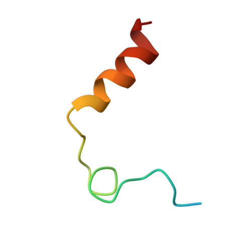
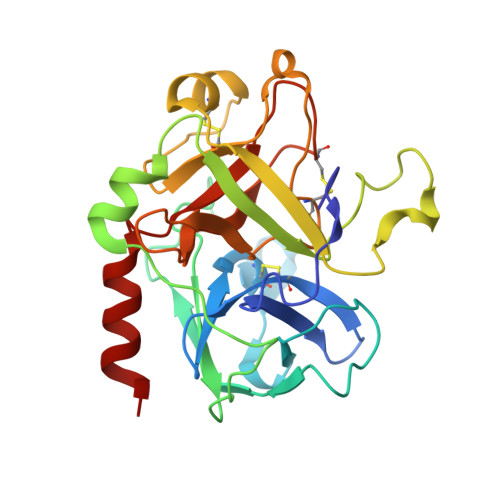
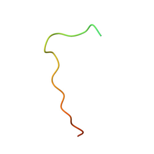
    The inhibition of thrombin is one of the important treatments of pathological blood clot formation. Variegin, isolated from the tropical bont tick, is a novel molecule exhibiting a unique 'two-modes' inhibitory property on thrombin active site (competitive before cleavage, noncompetitive after cleavage). For the better understanding of its function, we have determined the crystal structure of the human α-thrombin:synthetic-variegin complex at 2.4 Å resolution. The structure reveals a new mechanism of thrombin inhibition by disrupting the charge relay system. Based on the structure, we have designed 17 variegin variants, differing in potency, kinetics and mechanism of inhibition. The most active variant is about 70 times more potent than the FDA-approved peptidic thrombin inhibitor, hirulog-1/bivalirudin. In vivo antithrombotic effects of the variegin variants correlate well with their in vitro affinities for thrombin. Our results encourage that variegin and the variants show strong potential for the development of tunable anticoagulants.


  • Organizational Affiliation

    Department of Biological Sciences, National University of Singapore, Singapore, Singapore.


Macromolecules
Find similar proteins by:  (by identity cutoff)  |  3D Structure
Entity ID: 1
MoleculeChains Sequence LengthOrganismDetailsImage
Thrombin light chain36Homo sapiensMutation(s): 0 
Gene Names: F2
EC: 3.4.21.5
UniProt & NIH Common Fund Data Resources
Find proteins for P00734 (Homo sapiens)
Explore P00734 
Go to UniProtKB:  P00734
PHAROS:  P00734
GTEx:  ENSG00000180210 
Entity Groups  
Sequence Clusters30% Identity50% Identity70% Identity90% Identity95% Identity100% Identity
UniProt GroupP00734
Sequence Annotations
Expand
  • Reference Sequence
Find similar proteins by:  (by identity cutoff)  |  3D Structure
Entity ID: 2
MoleculeChains Sequence LengthOrganismDetailsImage
Thrombin heavy chain259Homo sapiensMutation(s): 0 
Gene Names: F2
EC: 3.4.21.5
UniProt & NIH Common Fund Data Resources
Find proteins for P00734 (Homo sapiens)
Explore P00734 
Go to UniProtKB:  P00734
PHAROS:  P00734
GTEx:  ENSG00000180210 
Entity Groups  
Sequence Clusters30% Identity50% Identity70% Identity90% Identity95% Identity100% Identity
UniProt GroupP00734
Sequence Annotations
Expand
  • Reference Sequence
Find similar proteins by:  (by identity cutoff)  |  3D Structure
Entity ID: 3
MoleculeChains Sequence LengthOrganismDetailsImage
Variegin32Amblyomma variegatumMutation(s): 0 
UniProt
Find proteins for P85800 (Amblyomma variegatum)
Explore P85800 
Go to UniProtKB:  P85800
Entity Groups  
Sequence Clusters30% Identity50% Identity70% Identity90% Identity95% Identity100% Identity
UniProt GroupP85800
Sequence Annotations
Expand
  • Reference Sequence
Experimental Data & Validation

Experimental Data

  • Method: X-RAY DIFFRACTION
  • Resolution: 2.40 Å
  • R-Value Free: 0.259 
  • R-Value Work: 0.208 
  • R-Value Observed: 0.210 
  • Space Group: C 1 2 1
Unit Cell:
Length ( Å )Angle ( ˚ )
a = 124.658α = 90
b = 50.826β = 98.7
c = 61.535γ = 90
Software Package:
Software NamePurpose
PHASERphasing
REFMACrefinement
HKL-2000data reduction
HKL-2000data scaling

Structure Validation

View Full Validation Report


Entry History 

Deposition Data

Revision History  (Full details and data files)

  • Version 1.0: 2011-11-23
    Type: Initial release
  • Version 1.1: 2023-11-01
    Changes: Data collection, Database references, Refinement description