3AYM

Crystal structure of the batho intermediate of squid rhodopsin


Experimental Data Snapshot

  • Method: X-RAY DIFFRACTION
  • Resolution: 2.80 Å
  • R-Value Free: 0.345 
  • R-Value Work: 0.295 

wwPDB Validation   3D Report Full Report


Ligand Structure Quality Assessment 


This is version 1.4 of the entry. See complete history


Literature

Crystallographic Analysis of the Primary Photochemical Reaction of Squid Rhodopsin

Murakami, M.Kouyama, T.

(2011) J Mol Biol 413: 615-627

  • DOI: https://doi.org/10.1016/j.jmb.2011.08.044
  • Primary Citation of Related Structures:  
    3AYM, 3AYN

  • PubMed Abstract: 

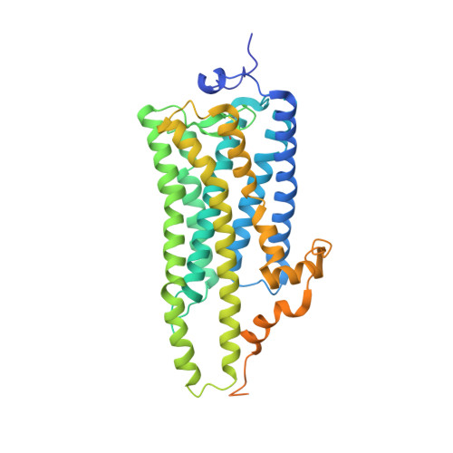
    Visual signal transduction is initiated by the photoisomerization of 11-cis retinal upon rhodopsin ligation. Unlike vertebrate rhodopsin, which interacts with Gt-type G-protein to stimulate the cyclic GMP signaling pathway, invertebrate rhodopsin interacts with Gq-type G-protein to stimulate a signaling pathway that is based on inositol 1,4,5-triphosphate. Since the inositol 1,4,5-triphosphate signaling pathway is utilized by mammalian nonvisual pigments and a large number of G-protein-coupled receptors, it is important to elucidate how the activation mechanism of invertebrate rhodopsin differs from that of vertebrate rhodopsin. Previous crystallographic studies of squid and bovine rhodopsins have shown that there is a profound difference in the structures of the retinal-binding pockets of these photoreceptors. Here, we report the crystal structures of all-trans bathorhodopsin (Batho; the first photoreaction intermediate) and the artificial 9-cis isorhodopsin (Iso) of squid rhodopsin. Upon the formation of Batho, the central moiety of the retinal was observed to move largely towards the cytoplasmic side, while the Schiff base and the ionone ring underwent limited movements (i.e., the all-trans retinal in Batho took on a right-handed screwed configuration). Conversely, the 9-cis retinal in Iso took on a planar configuration. Our results suggest that the light energy absorbed by squid rhodopsin is mostly converted into the distortion energy of the retinal polyene chain and surrounding residues.


  • Organizational Affiliation

    Department of Physics, Graduate School of Science, Nagoya University, Nagoya 464-8602, Japan.


Macromolecules
Find similar proteins by:  (by identity cutoff)  |  3D Structure
Entity ID: 1
MoleculeChains Sequence LengthOrganismDetailsImage
Rhodopsin
A, B
448Todarodes pacificusMutation(s): 0 
Membrane Entity: Yes 
UniProt
Find proteins for P31356 (Todarodes pacificus)
Explore P31356 
Go to UniProtKB:  P31356
Entity Groups  
Sequence Clusters30% Identity50% Identity70% Identity90% Identity95% Identity100% Identity
UniProt GroupP31356
Sequence Annotations
Expand
  • Reference Sequence
Small Molecules
Ligands 5 Unique
IDChains Name / Formula / InChI Key2D Diagram3D Interactions
PC1
Query on PC1

Download Ideal Coordinates CCD File 
G [auth B]1,2-DIACYL-SN-GLYCERO-3-PHOSPHOCHOLINE
C44 H88 N O8 P
NRJAVPSFFCBXDT-HUESYALOSA-N
BOG
Query on BOG

Download Ideal Coordinates CCD File 
D [auth A],
H [auth B]
octyl beta-D-glucopyranoside
C14 H28 O6
HEGSGKPQLMEBJL-RKQHYHRCSA-N
RET
Query on RET

Download Ideal Coordinates CCD File 
E [auth A],
I [auth B]
RETINAL
C20 H28 O
NCYCYZXNIZJOKI-OVSJKPMPSA-N
PLM
Query on PLM

Download Ideal Coordinates CCD File 
C [auth A]PALMITIC ACID
C16 H32 O2
IPCSVZSSVZVIGE-UHFFFAOYSA-N
SO4
Query on SO4

Download Ideal Coordinates CCD File 
F [auth B]SULFATE ION
O4 S
QAOWNCQODCNURD-UHFFFAOYSA-L
Experimental Data & Validation

Experimental Data

  • Method: X-RAY DIFFRACTION
  • Resolution: 2.80 Å
  • R-Value Free: 0.345 
  • R-Value Work: 0.295 
  • Space Group: P 62
Unit Cell:
Length ( Å )Angle ( ˚ )
a = 122.36α = 90
b = 122.36β = 90
c = 158.22γ = 120
Software Package:
Software NamePurpose
MAR345data collection
CNSrefinement
MOSFLMdata reduction
SCALAdata scaling
CNSphasing

Structure Validation

View Full Validation Report


Ligand Structure Quality Assessment 


Entry History 

Deposition Data

Revision History  (Full details and data files)

  • Version 1.0: 2011-08-17
    Type: Initial release
  • Version 1.1: 2011-10-26
    Changes: Database references
  • Version 1.2: 2017-10-11
    Changes: Refinement description
  • Version 1.3: 2020-07-29
    Type: Remediation
    Reason: Carbohydrate remediation
    Changes: Data collection, Derived calculations, Structure summary
  • Version 1.4: 2023-11-01
    Changes: Data collection, Database references, Refinement description, Structure summary