3AXB

Structure of a dye-linked L-proline dehydrogenase from the aerobic hyperthermophilic archaeon, Aeropyrum pernix


Experimental Data Snapshot

  • Method: X-RAY DIFFRACTION
  • Resolution: 1.92 Å
  • R-Value Free: 0.205 
  • R-Value Work: 0.188 
  • R-Value Observed: 0.189 

wwPDB Validation   3D Report Full Report


Ligand Structure Quality Assessment 


This is version 1.3 of the entry. See complete history


Literature

Crystal Structure of Novel Dye-linked L-Proline Dehydrogenase from Hyperthermophilic Archaeon Aeropyrum pernix

Sakuraba, H.Satomura, T.Kawakami, R.Kim, K.Hara, Y.Yoneda, K.Ohshima, T.

(2012) J Biol Chem 287: 20070-20080

  • DOI: https://doi.org/10.1074/jbc.M111.319038
  • Primary Citation of Related Structures:  
    3AXB, 3VQR

  • PubMed Abstract: 

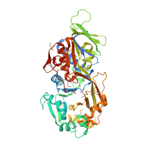
    Two types of dye-linked L-proline dehydrogenase (PDH1, α4β4-type hetero-octamer, and PDH2, αβγδ-type heterotetramer) have been identified so far in hyperthermophilic archaea. Here, we report the crystal structure of a third type of L-proline dehydrogenase, found in the aerobic hyperthermophilic archaeon Aeropyrum pernix, whose structure (homodimer) is much simpler than those of previously studied L-proline dehydrogenases. The structure was determined at a resolution of 1.92 Å. The asymmetric unit contained one subunit, and a crystallographic 2-fold axis generated the functional dimer. The overall fold of the subunit showed similarity to that of the PDH1 β-subunit, which is responsible for catalyzing L-proline dehydrogenation. However, the situation at the subunit-subunit interface of the A. pernix enzyme was totally different from that in PDH1. The presence of additional surface elements in the A. pernix enzyme contributes to a unique dimer association. Moreover, the C-terminal Leu(428), which is provided by a tail extending from the FAD-binding domain, shielded the active site, and an L-proline molecule was entrapped within the active site cavity. The K(m) value of a Leu(428) deletion mutant for L-proline was about 800 times larger than the K(m) value of the wild-type enzyme, although the k(cat) values did not differ much between the two enzymes. This suggests the C-terminal Leu(428) is not directly involved in catalysis, but it is essential for maintaining a high affinity for the substrate. This is the first description of an LPDH structure with L-proline bound, and it provides new insight into the substrate binding of LPDH.


  • Organizational Affiliation

    Department of Applied Biological Science, Faculty of Agriculture, Kagawa University, 2393 Ikenobe, Miki-cho, Kita-gun, Kagawa 761-0795, Japan.


Macromolecules
Find similar proteins by:  (by identity cutoff)  |  3D Structure
Entity ID: 1
MoleculeChains Sequence LengthOrganismDetailsImage
Putative oxidoreductase448Aeropyrum pernixMutation(s): 0 
UniProt
Find proteins for Q9YCJ0 (Aeropyrum pernix (strain ATCC 700893 / DSM 11879 / JCM 9820 / NBRC 100138 / K1))
Explore Q9YCJ0 
Go to UniProtKB:  Q9YCJ0
Entity Groups  
Sequence Clusters30% Identity50% Identity70% Identity90% Identity95% Identity100% Identity
UniProt GroupQ9YCJ0
Sequence Annotations
Expand
  • Reference Sequence
Small Molecules
Ligands 3 Unique
IDChains Name / Formula / InChI Key2D Diagram3D Interactions
FAD
Query on FAD

Download Ideal Coordinates CCD File 
C [auth A]FLAVIN-ADENINE DINUCLEOTIDE
C27 H33 N9 O15 P2
VWWQXMAJTJZDQX-UYBVJOGSSA-N
PRO
Query on PRO

Download Ideal Coordinates CCD File 
B [auth A]PROLINE
C5 H9 N O2
ONIBWKKTOPOVIA-BYPYZUCNSA-N
EDO
Query on EDO

Download Ideal Coordinates CCD File 
D [auth A]
E [auth A]
F [auth A]
G [auth A]
H [auth A]
D [auth A],
E [auth A],
F [auth A],
G [auth A],
H [auth A],
I [auth A]
1,2-ETHANEDIOL
C2 H6 O2
LYCAIKOWRPUZTN-UHFFFAOYSA-N
Modified Residues  1 Unique
IDChains TypeFormula2D DiagramParent
MSE
Query on MSE
A
L-PEPTIDE LINKINGC5 H11 N O2 SeMET
Experimental Data & Validation

Experimental Data

  • Method: X-RAY DIFFRACTION
  • Resolution: 1.92 Å
  • R-Value Free: 0.205 
  • R-Value Work: 0.188 
  • R-Value Observed: 0.189 
  • Space Group: P 43 21 2
Unit Cell:
Length ( Å )Angle ( ˚ )
a = 60.182α = 90
b = 60.182β = 90
c = 274.096γ = 90
Software Package:
Software NamePurpose
DENZOdata reduction
SCALEPACKdata scaling
SOLVEphasing
RESOLVEphasing
REFMACrefinement
PDB_EXTRACTdata extraction
HKL-2000data collection

Structure Validation

View Full Validation Report


Ligand Structure Quality Assessment 


Entry History 

Deposition Data

Revision History  (Full details and data files)

  • Version 1.0: 2012-04-04
    Type: Initial release
  • Version 1.1: 2012-04-25
    Changes: Database references
  • Version 1.2: 2012-06-20
    Changes: Database references
  • Version 1.3: 2017-10-11
    Changes: Refinement description