3AX5

Crystal structure of rat TOM20-ALDH presequence complex: A complex (form1) between Tom20 and a disulfide-bridged presequence peptide containing D-Cys and L-Cys at the i and i+3 positions.


Experimental Data Snapshot

  • Method: X-RAY DIFFRACTION
  • Resolution: 2.20 Å
  • R-Value Free: 0.276 
  • R-Value Work: 0.222 
  • R-Value Observed: 0.225 

wwPDB Validation   3D Report Full Report


This is version 1.2 of the entry. See complete history


Literature

Crystallographic snapshots of tom20-mitochondrial presequence interactions with disulfide-stabilized peptides.

Saitoh, T.Igura, M.Miyazaki, Y.Ose, T.Maita, N.Kohda, D.

(2011) Biochemistry 50: 5487-5496

  • DOI: https://doi.org/10.1021/bi200470x
  • Primary Citation of Related Structures:  
    3AWR, 3AX2, 3AX3, 3AX5

  • PubMed Abstract: 

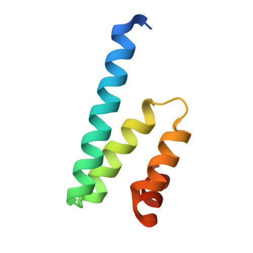
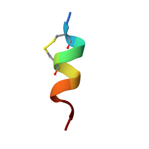
    Most mitochondrial proteins are synthesized in the cytosol and imported into mitochondria. The Tom20 protein, residing on the mitochondrial surface, recognizes the N-terminal presequences of precursor proteins. We previously determined the crystal structures of the Tom20-presequence complex. The successful crystallization involved tethering the presequence to Tom20 through an intermolecular disulfide bond with an optimized linker. In this work, we assessed the tethering method. The intermolecular disulfide bond was cleaved in crystal with a reducing agent. The pose (i.e., conformation and position) of the presequence was identical to the previously determined pose. In another experiment, a longer linker than the optimized length was used for the tethering. The perturbation of the tether changed the pose slightly, but the interaction mode was preserved. These results argue against the forced interaction of the presequence by its covalent attachment to Tom20. Second, as an alternative method referred to as "molecular stiffening", we introduced a disulfide bond within the presequence peptide to restrict the freedom of the peptide in the unbound states. One presequence analogue exhibited over 100-fold higher affinity than its linear counterpart and generated cocrystals with Tom20. One of the two crystallographic snapshots revealed a known pose previously determined by the tethering method, and the other snapshot depicted a new pose. These results confirmed and extended the dynamic, multiple bound state model of the Tom20-presequence interactions and also demonstrated the validity of the molecular tethering and stiffening techniques in studies of transient protein-peptide interactions.


  • Organizational Affiliation

    Division of Structural Biology, Medical Institute of Bioregulation, Kyushu University, Maidashi 3-1-1, Higashi-ku, Fukuoka 812-8582, Japan.


Macromolecules
Find similar proteins by:  (by identity cutoff)  |  3D Structure
Entity ID: 1
MoleculeChains Sequence LengthOrganismDetailsImage
Mitochondrial import receptor subunit TOM20 homolog
A, C
73Rattus norvegicusMutation(s): 0 
Gene Names: Tomm20
UniProt
Find proteins for Q62760 (Rattus norvegicus)
Explore Q62760 
Go to UniProtKB:  Q62760
Entity Groups  
Sequence Clusters30% Identity50% Identity70% Identity90% Identity95% Identity100% Identity
UniProt GroupQ62760
Sequence Annotations
Expand
  • Reference Sequence

Find similar proteins by:  Sequence   |   3D Structure  

Entity ID: 2
MoleculeChains Sequence LengthOrganismDetailsImage
Aldehyde dehydrogenase, mitochondrial
B, D
11Rattus norvegicusMutation(s): 2 
UniProt
Find proteins for P11884 (Rattus norvegicus)
Explore P11884 
Go to UniProtKB:  P11884
Entity Groups  
Sequence Clusters30% Identity50% Identity70% Identity90% Identity95% Identity100% Identity
UniProt GroupP11884
Sequence Annotations
Expand
  • Reference Sequence
Small Molecules
Ligands 1 Unique
IDChains Name / Formula / InChI Key2D Diagram3D Interactions
PO4
Query on PO4

Download Ideal Coordinates CCD File 
E [auth A]PHOSPHATE ION
O4 P
NBIIXXVUZAFLBC-UHFFFAOYSA-K
Experimental Data & Validation

Experimental Data

  • Method: X-RAY DIFFRACTION
  • Resolution: 2.20 Å
  • R-Value Free: 0.276 
  • R-Value Work: 0.222 
  • R-Value Observed: 0.225 
  • Space Group: I 2 2 2
Unit Cell:
Length ( Å )Angle ( ˚ )
a = 41.745α = 90
b = 77.401β = 90
c = 116.226γ = 90
Software Package:
Software NamePurpose
PHASERphasing
REFMACrefinement
HKL-2000data reduction
HKL-2000data scaling

Structure Validation

View Full Validation Report


Entry History 

Deposition Data

Revision History  (Full details and data files)

  • Version 1.0: 2011-07-06
    Type: Initial release
  • Version 1.1: 2011-07-13
    Changes: Version format compliance
  • Version 1.2: 2023-11-01
    Changes: Data collection, Database references, Derived calculations, Refinement description