


Structure of UDP-galactose 4-epimerase mutant
Sakuraba, H., Kawai, T., Yoneda, K., Ohshima, T.To be published.
Experimental Data Snapshot
Entity ID: 1 | |||||
|---|---|---|---|---|---|
| Molecule | Chains | Sequence Length | Organism | Details | Image |
| NAD-dependent epimerase/dehydratase | 308 | Pyrobaculum calidifontis JCM 11548 | Mutation(s): 0 Gene Names: Pcal_0885 | 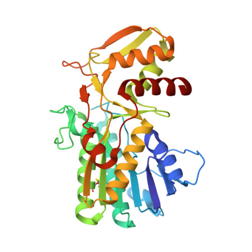 | |
UniProt | |||||
Find proteins for A3MUJ4 (Pyrobaculum calidifontis (strain DSM 21063 / JCM 11548 / VA1)) Explore A3MUJ4 Go to UniProtKB: A3MUJ4 | |||||
Entity Groups | |||||
| Sequence Clusters | 30% Identity50% Identity70% Identity90% Identity95% Identity100% Identity | ||||
| UniProt Group | A3MUJ4 | ||||
Sequence AnnotationsExpand | |||||
| |||||
| Ligands 2 Unique | |||||
|---|---|---|---|---|---|
| ID | Chains | Name / Formula / InChI Key | 2D Diagram | 3D Interactions | |
| NAD Query on NAD | E [auth A], G [auth B], I [auth C] | NICOTINAMIDE-ADENINE-DINUCLEOTIDE C21 H27 N7 O14 P2 BAWFJGJZGIEFAR-NNYOXOHSSA-N |  | ||
| GDU Query on GDU | D [auth A], F [auth B], H [auth C] | GALACTOSE-URIDINE-5'-DIPHOSPHATE C15 H24 N2 O17 P2 HSCJRCZFDFQWRP-ABVWGUQPSA-N |  | ||
| Length ( Å ) | Angle ( ˚ ) |
|---|---|
| a = 88.623 | α = 90 |
| b = 113.644 | β = 90 |
| c = 217.267 | γ = 90 |
| Software Name | Purpose |
|---|---|
| DENZO | data reduction |
| SCALEPACK | data scaling |
| MOLREP | phasing |
| DM | phasing |
| REFMAC | refinement |
| PDB_EXTRACT | data extraction |
| HKL-2000 | data collection |
| HKL-2000 | data reduction |
| HKL-2000 | data scaling |
RCSB PDB (citation) is hosted by
RCSB PDB is a member of the

