Crystal structure of bacterial cell-surface alginate-binding protein with an M75 peptidase motif.
Maruyama, Y., Ochiai, A., Mikami, B., Hashimoto, W., Murata, K.(2011) Biochem Biophys Res Commun 405: 411-416
- PubMed: 21238429
- DOI: https://doi.org/10.1016/j.bbrc.2011.01.043
- Primary Citation of Related Structures:
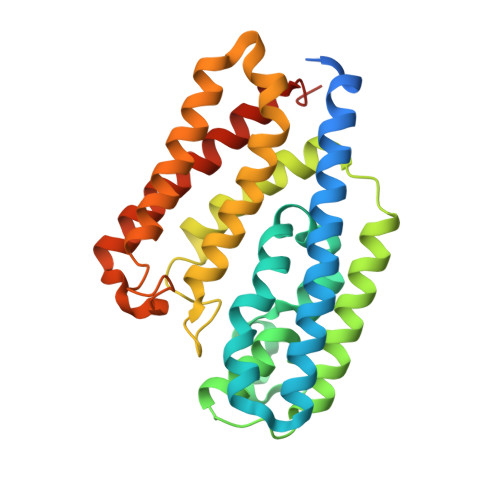
3AT7 - PubMed Abstract:
A gram-negative Sphingomonas sp. A1 directly incorporates alginate polysaccharide into the cytoplasm via the cell-surface pit and ABC transporter. A cell-surface alginate-binding protein, Algp7, functions as a concentrator of the polysaccharide in the pit. Based on the primary structure and genetic organization in the bacterial genome, Algp7 was found to be homologous to an M75 peptidase motif-containing EfeO, a component of a ferrous ion transporter. Despite the presence of an M75 peptidase motif with high similarity, the Algp7 protein purified from recombinant Escherichia coli cells was inert on insulin B chain and N-benzoyl-Phe-Val-Arg-p-nitroanilide, both of which are substrates for a typical M75 peptidase, imelysin, from Pseudomonas aeruginosa. The X-ray crystallographic structure of Algp7 was determined at 2.10Å resolution by single-wavelength anomalous diffraction. Although a metal-binding motif, HxxE, conserved in zinc ion-dependent M75 peptidases is also found in Algp7, the crystal structure of Algp7 contains no metal even at the motif. The protein consists of two structurally similar up-and-down helical bundles as the basic scaffold. A deep cleft between the bundles is sufficiently large to accommodate macromolecules such as alginate polysaccharide. This is the first structural report on a bacterial cell-surface alginate-binding protein with an M75 peptidase motif.
Organizational Affiliation:
Laboratory of Basic and Applied Molecular Biotechnology, Graduate School of Agriculture, Kyoto University, Uji, Kyoto 611-0011, Japan.