3AQA

Crystal structure of the human BRD2 BD1 bromodomain in complex with a BRD2-interactive compound, BIC1


Experimental Data Snapshot

  • Method: X-RAY DIFFRACTION
  • Resolution: 2.30 Å
  • R-Value Free: 0.245 
  • R-Value Work: 0.189 
  • R-Value Observed: 0.189 

wwPDB Validation   3D Report Full Report


Ligand Structure Quality Assessment 


This is version 1.3 of the entry. See complete history


Literature

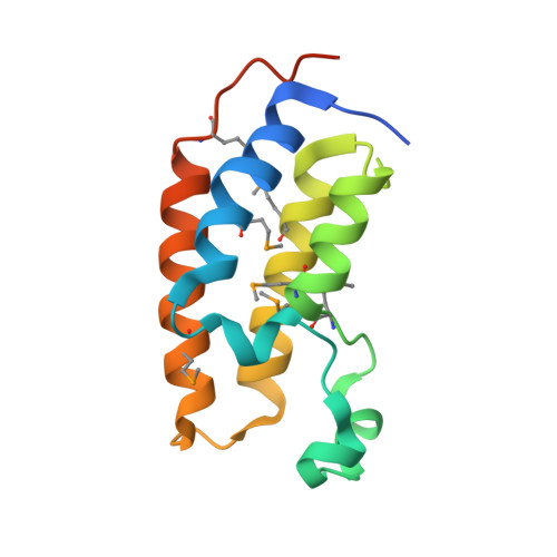
Real-Time Imaging of Histone H4K12-Specific Acetylation Determines the Modes of Action of Histone Deacetylase and Bromodomain Inhibitors

Ito, T.Umehara, T.Sasaki, K.Nakamura, Y.Nishino, N.Terada, T.Shirouzu, M.Padmanabhan, B.Yokoyama, S.Ito, A.Yoshida, M.

(2011) Chem Biol 18: 495-507

  • DOI: https://doi.org/10.1016/j.chembiol.2011.02.009
  • Primary Citation of Related Structures:  
    3AQA

  • PubMed Abstract: 

    Histone acetylation constitutes an epigenetic mark for transcriptional regulation. Here we developed a fluorescent probe to visualize acetylation of histone H4 Lys12 (H4K12) in living cells using fluorescence resonance energy transfer (FRET) and the binding of the BRD2 bromodomain to acetylated H4K12. Using this probe designated as Histac-K12, we demonstrated that histone H4K12 acetylation is retained in mitosis and that some histone deacetylase (HDAC) inhibitors continue to inhibit cellular HDAC activity even after their removal from the culture. In addition, a small molecule that interferes with ability of the bromodomain to bind to acetylated H4K12 could be assessed using Histac-K12 in cells. Thus, Histac-K12 will serve as a powerful tool not only to understand the dynamics of H4K12-specific acetylation but also to characterize small molecules that modulate the acetylation or interaction status of histones.


  • Organizational Affiliation

    Chemical Genetics Laboratory, RIKEN Advanced Science Institute, Wako, Saitama 351-0198, Japan.


Macromolecules
Find similar proteins by:  (by identity cutoff)  |  3D Structure
Entity ID: 1
MoleculeChains Sequence LengthOrganismDetailsImage
Bromodomain-containing protein 2
A, B, C
128Homo sapiensMutation(s): 0 
Gene Names: BRD2KIAA9001RING3
UniProt & NIH Common Fund Data Resources
Find proteins for P25440 (Homo sapiens)
Explore P25440 
Go to UniProtKB:  P25440
PHAROS:  P25440
GTEx:  ENSG00000204256 
Entity Groups  
Sequence Clusters30% Identity50% Identity70% Identity90% Identity95% Identity100% Identity
UniProt GroupP25440
Sequence Annotations
Expand
  • Reference Sequence
Small Molecules
Ligands 2 Unique
IDChains Name / Formula / InChI Key2D Diagram3D Interactions
BYH
Query on BYH

Download Ideal Coordinates CCD File 
D [auth A]1-[2-(1H-benzimidazol-2-ylsulfanyl)ethyl]-3-methyl-1,3-dihydro-2H-benzimidazole-2-thione
C17 H16 N4 S2
KDPSGIFCBZTBEZ-UHFFFAOYSA-N
MES
Query on MES

Download Ideal Coordinates CCD File 
E [auth A]2-(N-MORPHOLINO)-ETHANESULFONIC ACID
C6 H13 N O4 S
SXGZJKUKBWWHRA-UHFFFAOYSA-N
Modified Residues  1 Unique
IDChains TypeFormula2D DiagramParent
MSE
Query on MSE
A, B, C
L-PEPTIDE LINKINGC5 H11 N O2 SeMET
Binding Affinity Annotations 
IDSourceBinding Affinity
BYH PDBBind:  3AQA Kd: 2.80e+4 (nM) from 1 assay(s)
Experimental Data & Validation

Experimental Data

  • Method: X-RAY DIFFRACTION
  • Resolution: 2.30 Å
  • R-Value Free: 0.245 
  • R-Value Work: 0.189 
  • R-Value Observed: 0.189 
  • Space Group: C 1 2 1
Unit Cell:
Length ( Å )Angle ( ˚ )
a = 114.597α = 90
b = 55.716β = 94.17
c = 68.205γ = 90
Software Package:
Software NamePurpose
CrystalCleardata collection
MOLREPphasing
CNSrefinement
HKL-2000data reduction
HKL-2000data scaling

Structure Validation

View Full Validation Report


Ligand Structure Quality Assessment 


Entry History 

Revision History  (Full details and data files)

  • Version 1.0: 2011-05-18
    Type: Initial release
  • Version 1.1: 2011-07-13
    Changes: Version format compliance
  • Version 1.2: 2023-11-01
    Changes: Data collection, Database references, Derived calculations, Refinement description
  • Version 1.3: 2023-11-15
    Changes: Data collection