3AJW

Structure of FliJ, a soluble component of flagellar type III export apparatus


Experimental Data Snapshot

  • Method: X-RAY DIFFRACTION
  • Resolution: 2.10 Å
  • R-Value Free: 0.255 
  • R-Value Work: 0.232 
  • R-Value Observed: 0.232 

wwPDB Validation   3D Report Full Report


This is version 1.2 of the entry. See complete history


Literature

Common architecture of the flagellar type III protein export apparatus and F- and V-type ATPases

Ibuki, T.Imada, K.Minamino, T.Kato, T.Miyata, T.Namba, K.

(2011) Nat Struct Mol Biol 18: 277-282

  • DOI: https://doi.org/10.1038/nsmb.1977
  • Primary Citation of Related Structures:  
    3AJW

  • PubMed Abstract: 

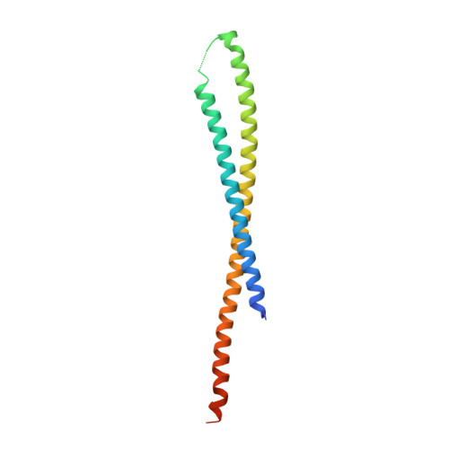
    The proteins that form the bacterial flagellum are translocated to its distal end through the central channel of the growing flagellum by the flagellar-specific protein export apparatus, a family of the type III protein secretion system. FliI and FliJ are soluble components of this apparatus. FliI is an ATPase that has extensive structural similarity to the α and β subunits of F(o)F(1)-ATP synthase. FliJ is essential for export, but its function remains obscure. Here we show that the structure of FliJ derived from Salmonella enterica serovar Typhimurium is remarkably similar to that of the two-stranded α-helical coiled-coil part of the γ subunit of F(o)F(1)-ATP synthase and that FliJ promotes the formation of FliI hexamer rings by binding to the center of the ring. These results suggest that the type III protein export system and F- and V-type ATPases share a similar mechanism and an evolutionary relationship.


  • Organizational Affiliation

    Osaka University, Osaka, Japan.


Macromolecules
Find similar proteins by:  (by identity cutoff)  |  3D Structure
Entity ID: 1
MoleculeChains Sequence LengthOrganismDetailsImage
Flagellar fliJ protein150Salmonella enterica subsp. enterica serovar TyphimuriumMutation(s): 0 
UniProt
Find proteins for P0A1K1 (Salmonella typhimurium (strain LT2 / SGSC1412 / ATCC 700720))
Explore P0A1K1 
Go to UniProtKB:  P0A1K1
Entity Groups  
Sequence Clusters30% Identity50% Identity70% Identity90% Identity95% Identity100% Identity
UniProt GroupP0A1K1
Sequence Annotations
Expand
  • Reference Sequence
Small Molecules
Ligands 1 Unique
IDChains Name / Formula / InChI Key2D Diagram3D Interactions
HG
Query on HG

Download Ideal Coordinates CCD File 
B [auth A]MERCURY (II) ION
Hg
BQPIGGFYSBELGY-UHFFFAOYSA-N
Experimental Data & Validation

Experimental Data

  • Method: X-RAY DIFFRACTION
  • Resolution: 2.10 Å
  • R-Value Free: 0.255 
  • R-Value Work: 0.232 
  • R-Value Observed: 0.232 
  • Space Group: P 65 2 2
Unit Cell:
Length ( Å )Angle ( ˚ )
a = 52.925α = 90
b = 52.925β = 90
c = 192.597γ = 120
Software Package:
Software NamePurpose
ADSCdata collection
SOLVEphasing
CNSrefinement
MOSFLMdata reduction
SCALAdata scaling

Structure Validation

View Full Validation Report


Entry History 

Deposition Data

Revision History  (Full details and data files)

  • Version 1.0: 2011-02-02
    Type: Initial release
  • Version 1.1: 2011-07-13
    Changes: Version format compliance
  • Version 1.2: 2024-03-13
    Changes: Data collection, Database references, Derived calculations