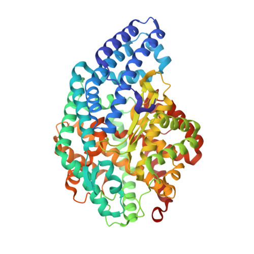
3AHN

PZ PEPTIDASE A with Inhibitor 1


Experimental Data Snapshot

  • Method: X-RAY DIFFRACTION
  • Resolution: 1.80 Å
  • R-Value Free: 0.200 
  • R-Value Work: 0.188 
  • R-Value Observed: 0.189 

wwPDB Validation   3D Report Full Report


Ligand Structure Quality Assessment 


This is version 1.4 of the entry. See complete history


Literature

The exquisite structure and reaction mechanism of bacterial Pz-peptidase A toward collagenous peptides: X-ray crystallographic structure analysis of PZ-peptidase a reveals differences from mammalian thimet oligopeptidase.

Kawasaki, A.Nakano, H.Hosokawa, A.Nakatsu, T.Kato, H.Watanabe, K.

(2010) J Biol Chem 285: 34972-34980

  • DOI: https://doi.org/10.1074/jbc.M110.141838
  • Primary Citation of Related Structures:  
    3AHM, 3AHN, 3AHO

  • PubMed Abstract: 

    Pz-peptidase A, from the thermophilic bacterium Geobacillus collagenovorans MO-1, hydrolyzes a synthetic peptide substrate, 4-phenylazobenzyloxycarbonyl-Pro-Leu-Gly-Pro-D-Arg (Pz-PLGPR), which contains a collagen-specific tripeptide sequence, -Gly-Pro-X-, but does not act on collagen proteins themselves. The mammalian enzyme, thimet oligopeptidase (TOP), which has comparable functions with bacterial Pz-peptidases but limited identity at the primary sequence level, has recently been subjected to x-ray crystallographic analysis; however, no crystal structure has yet been reported for complexes of TOP with substrate analogues. Here, we report crystallization of recombinant Pz-peptidase A in complex with two phosphinic peptide inhibitors (PPIs) that also function as inhibitors of TOP and determination of the crystal structure of these complexes at 1.80-2.00 Å resolution. The most striking difference between Pz-peptidase A and TOP is that there is no channel running the length of bacterial protein. Whereas the structure of TOP resembles an open bivalve, that of Pz-peptidase A is closed and globular. This suggests that collagenous peptide substrates enter the tunnel at the top gateway of the closed Pz-peptidase A molecule, and reactant peptides are released from the bottom gateway after cleavage at the active site located in the center of the tunnel. One of the two PPIs, PPI-2, which contains the collagen-specific sequence, helped to clarify the exquisite structure and reaction mechanism of Pz-peptidase A toward collagenous peptides. This study describes the mode of substrate binding and its implication for the mammalian enzymes.


  • Organizational Affiliation

    From the Division of Applied Life Sciences, Graduate School of Life and Environmental Sciences, Kyoto Prefectural University, Shimogamo, Sakyo, Kyoto 606-8522, Japan.


Macromolecules
Find similar proteins by:  (by identity cutoff)  |  3D Structure
Entity ID: 1
MoleculeChains Sequence LengthOrganismDetailsImage
Oligopeptidase
A, B
564Geobacillus sp. MO-1Mutation(s): 0 
UniProt
Find proteins for Q4W803 (Geobacillus sp. MO-1)
Explore Q4W803 
Go to UniProtKB:  Q4W803
Entity Groups  
Sequence Clusters30% Identity50% Identity70% Identity90% Identity95% Identity100% Identity
UniProt GroupQ4W803
Sequence Annotations
Expand
  • Reference Sequence
Small Molecules
Ligands 3 Unique
IDChains Name / Formula / InChI Key2D Diagram3D Interactions
3A1
Query on 3A1

Download Ideal Coordinates CCD File 
C [auth A],
H [auth B]
N~2~-{(2S)-3-[(R)-hydroxy{(1R)-2-phenyl-1-[(phenylacetyl)amino]ethyl}phosphoryl]-2-methylpropanoyl}-L-lysyl-D-serine
C29 H41 N4 O8 P
CUHSSJWAGOQFDP-JOXZGHKQSA-N
ZN
Query on ZN

Download Ideal Coordinates CCD File 
D [auth A],
G [auth B]
ZINC ION
Zn
PTFCDOFLOPIGGS-UHFFFAOYSA-N
ACT
Query on ACT

Download Ideal Coordinates CCD File 
E [auth A],
F [auth A],
I [auth B],
J [auth B],
K [auth B]
ACETATE ION
C2 H3 O2
QTBSBXVTEAMEQO-UHFFFAOYSA-M
Binding Affinity Annotations 
IDSourceBinding Affinity
3A1 Binding MOAD:  3AHN Ki: 90.1 (nM) from 1 assay(s)
PDBBind:  3AHN Ki: 90.1 (nM) from 1 assay(s)
Experimental Data & Validation

Experimental Data

  • Method: X-RAY DIFFRACTION
  • Resolution: 1.80 Å
  • R-Value Free: 0.200 
  • R-Value Work: 0.188 
  • R-Value Observed: 0.189 
  • Space Group: P 1 21 1
Unit Cell:
Length ( Å )Angle ( ˚ )
a = 56.379α = 90
b = 194.148β = 106.22
c = 59.924γ = 90
Software Package:
Software NamePurpose
CrystalCleardata collection
MOLREPphasing
REFMACrefinement
MOSFLMdata reduction
SCALAdata scaling

Structure Validation

View Full Validation Report


Ligand Structure Quality Assessment 


Entry History 

Deposition Data

  • Released Date: 2010-08-25 
  • Deposition Author(s): Nakano, H.

Revision History  (Full details and data files)

  • Version 1.0: 2010-08-25
    Type: Initial release
  • Version 1.1: 2011-07-13
    Changes: Atomic model, Database references, Derived calculations, Non-polymer description, Structure summary, Version format compliance
  • Version 1.2: 2012-12-12
    Changes: Other
  • Version 1.3: 2015-12-16
    Changes: Database references
  • Version 1.4: 2024-02-21
    Changes: Data collection, Database references, Derived calculations