3AGP

Structure of viral polymerase form I


Experimental Data Snapshot

  • Method: X-RAY DIFFRACTION
  • Resolution: 2.80 Å
  • R-Value Free: 0.283 
  • R-Value Work: 0.215 

wwPDB Validation   3D Report Full Report


This is version 1.4 of the entry. See complete history


Literature

Assembly of Q{beta} viral RNA polymerase with host translational elongation factors EF-Tu and -Ts

Takeshita, D.Tomita, K.

(2010) Proc Natl Acad Sci U S A 107: 15733-15738

  • DOI: https://doi.org/10.1073/pnas.1006559107
  • Primary Citation of Related Structures:  
    3AGP, 3AGQ

  • PubMed Abstract: 

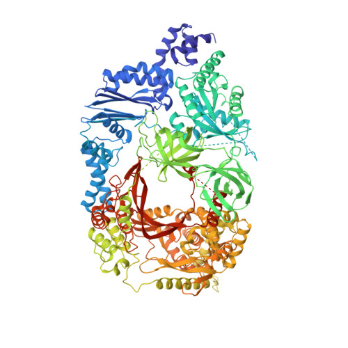
    Replication and transcription of viral RNA genomes rely on host-donated proteins. Qbeta virus infects Escherichia coli and replicates and transcribes its own genomic RNA by Qbeta replicase. Qbeta replicase requires the virus-encoded RNA-dependent RNA polymerase (beta-subunit), and the host-donated translational elongation factors EF-Tu and -Ts, as active core subunits for its RNA polymerization activity. Here, we present the crystal structure of the core Qbeta replicase, comprising the beta-subunit, EF-Tu and -Ts. The beta-subunit has a right-handed structure, and the EF-Tu:Ts binary complex maintains the structure of the catalytic core crevasse of the beta-subunit through hydrophobic interactions, between the finger and thumb domains of the beta-subunit and domain-2 of EF-Tu and the coiled-coil motif of EF-Ts, respectively. These hydrophobic interactions are required for the expression and assembly of the Qbeta replicase complex. Thus, EF-Tu and -Ts have chaperone-like functions in the maintenance of the structure of the active Qbeta replicase. Modeling of the template RNA and the growing RNA in the catalytic site of the Qbeta replicase structure also suggests that structural changes of the RNAs and EF-Tu:Ts should accompany processive RNA polymerization and that EF-Tu:Ts in the Qbeta replicase could function to modulate the RNA folding and structure.


  • Organizational Affiliation

    Biomedical Research Institute, National Institute of Advanced Industrial Science and Technology, Higashi, Tsukuba, Ibaraki, Japan.


Macromolecules
Find similar proteins by:  (by identity cutoff)  |  3D Structure
Entity ID: 1
MoleculeChains Sequence LengthOrganismDetailsImage
Elongation factor Ts, Elongation factor Tu, LINKER, Q beta replicase1,289Escherichia coli O157:H7synthetic constructQubevirus durumMutation(s): 0 
UniProt
Find proteins for P14647 (Escherichia virus Qbeta)
Explore P14647 
Go to UniProtKB:  P14647
Find proteins for P0A6N3 (Escherichia coli O157:H7)
Explore P0A6N3 
Go to UniProtKB:  P0A6N3
Find proteins for P0A6P3 (Escherichia coli O157:H7)
Explore P0A6P3 
Go to UniProtKB:  P0A6P3
Entity Groups  
Sequence Clusters30% Identity50% Identity70% Identity90% Identity95% Identity100% Identity
UniProt GroupsP14647P0A6P3P0A6N3
Sequence Annotations
Expand
  • Reference Sequence
Small Molecules
Ligands 1 Unique
IDChains Name / Formula / InChI Key2D Diagram3D Interactions
CA
Query on CA

Download Ideal Coordinates CCD File 
B [auth A]CALCIUM ION
Ca
BHPQYMZQTOCNFJ-UHFFFAOYSA-N
Experimental Data & Validation

Experimental Data

  • Method: X-RAY DIFFRACTION
  • Resolution: 2.80 Å
  • R-Value Free: 0.283 
  • R-Value Work: 0.215 
  • Space Group: C 2 2 21
Unit Cell:
Length ( Å )Angle ( ˚ )
a = 139.498α = 90
b = 256.428β = 90
c = 100.961γ = 90
Software Package:
Software NamePurpose
HKL-2000data collection
SnBphasing
PHENIXrefinement
HKL-2000data reduction
HKL-2000data scaling

Structure Validation

View Full Validation Report


Entry History 

Deposition Data

Revision History  (Full details and data files)

  • Version 1.0: 2010-09-01
    Type: Initial release
  • Version 1.1: 2011-07-13
    Changes: Version format compliance
  • Version 1.2: 2014-01-29
    Changes: Database references
  • Version 1.3: 2017-06-28
    Changes: Source and taxonomy
  • Version 1.4: 2024-03-13
    Changes: Data collection, Database references, Derived calculations