Molecular identification of unsaturated uronate reductase prerequisite for alginate metabolism in Sphingomonas sp. A1
Takase, R., Ochiai, A., Mikami, B., Hashimoto, W., Murata, K.(2010) Biochim Biophys Acta 1804: 1925-1936
- PubMed: 20685299
- DOI: https://doi.org/10.1016/j.bbapap.2010.05.010
- Primary Citation of Related Structures:
3AFM, 3AFN - PubMed Abstract:
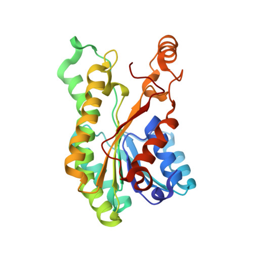
In Sphingomonas sp. A1, alginate is degraded by alginate lyases to its constituent monosaccharides, which are nonenzymatically converted to an alpha-keto acid, namely, 4-deoxy-l-erythro-5-hexoseulose uronic acid (DEH). The properties of the DEH-metabolizing enzyme and its gene in strain A1 were characterized. In the presence of alginate, strain A1 cells inducibly produced an NADPH-dependent DEH reductase (A1-R) in their cytoplasm. Molecular cloning of the enzyme gene indicated that A1-R belonged to the short-chain dehydrogenase/reductase superfamily and catalyzed the conversion of DEH to 2-keto-3-deoxy-d-gluconic acid most efficiently at around pH 7.0 and 50 degrees C. Crystal structures of A1-R and its complex with NADP were determined at around 1.6A resolution by X-ray crystallography. The enzyme consists of three layers (alpha/beta/alpha), with a coenzyme-binding Rossmann fold. NADP is surrounded by positively charged residues, and Gly-38 and Arg-39 are crucial for NADP binding. Site-directed mutagenesis studies suggest that Ser-150, Tyr-164, and Lys-168 located around the Rossmann fold constitute the catalytic triad. To our knowledge, this is the first report on molecular cloning and structure determination of a bacterial DEH reductase responsible for alginate metabolism.
Organizational Affiliation:
Laboratory of Basic and Applied Molecular Biotechnology, Graduate School of Agriculture, Kyoto University, Uji, Kyoto 611-0011, Japan.