3A8T

Plant adenylate isopentenyltransferase in complex with ATP


Experimental Data Snapshot

  • Method: X-RAY DIFFRACTION
  • Resolution: 2.37 Å
  • R-Value Free: 0.235 
  • R-Value Work: 0.181 
  • R-Value Observed: 0.184 

wwPDB Validation   3D Report Full Report


Ligand Structure Quality Assessment 


This is version 1.3 of the entry. See complete history


Literature

Crystal structure and substrate specificity of plant adenylate isopentenyltransferase from Humulus lupulus: distinctive binding affinity for purine and pyrimidine nucleotides

Chu, H.-M.Ko, T.-P.Wang, A.H.-J.

(2010) Nucleic Acids Res 38: 1738-1748

  • DOI: https://doi.org/10.1093/nar/gkp1093
  • Primary Citation of Related Structures:  
    3A8T

  • PubMed Abstract: 

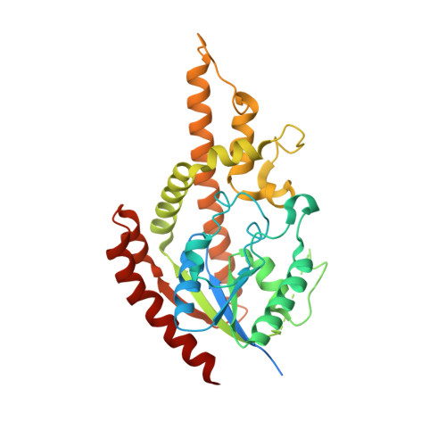
    Cytokinins are important plant hormones, and their biosynthesis most begins with the transfer of isopentenyl group from dimethylallyl diphosphate (DMAPP) to the N6-amino group of adenine by either adenylate isopentenyltransferase (AIPT) or tRNA-IPT. Plant AIPTs use ATP/ADP as an isopentenyl acceptor and bacterial AIPTs prefer AMP, whereas tRNA-IPTs act on specific sites of tRNA. Here, we present the crystal structure of an AIPT-ATP complex from Humulus lupulus (HlAIPT), which is similar to the previous structures of Agrobacterium AIPT and yeast tRNA-IPT. The enzyme is structurally homologous to the NTP-binding kinase family of proteins but forms a solvent-accessible channel that binds to the donor substrate DMAPP, which is directed toward the acceptor substrate ATP/ADP. When measured with isothermal titration calorimetry, some nucleotides displayed different binding affinities to HlAIPT with an order of ATP > dATP approximately ADP > GTP > CTP > UTP. Two basic residues Lys275 and Lys220 in HlAIPT interact with the beta and gamma-phosphate of ATP. By contrast, the interactions are absent in Agrobacterium AIPT because they are replaced by the acidic residues Asp221 and Asp171. Despite its structural similarity to the yeast tRNA-IPT, HlAIPT has evolved with a different binding strategy for adenylate.


  • Organizational Affiliation

    Institute of Biochemical Sciences, National Taiwan University, Taipei, 106, Taiwan.


Macromolecules
Find similar proteins by:  (by identity cutoff)  |  3D Structure
Entity ID: 1
MoleculeChains Sequence LengthOrganismDetailsImage
Adenylate isopentenyltransferase339Humulus lupulusMutation(s): 0 
Gene Names: AIPT
EC: 2.5.1.27
UniProt
Find proteins for Q5GHF7 (Humulus lupulus)
Explore Q5GHF7 
Go to UniProtKB:  Q5GHF7
Entity Groups  
Sequence Clusters30% Identity50% Identity70% Identity90% Identity95% Identity100% Identity
UniProt GroupQ5GHF7
Sequence Annotations
Expand
  • Reference Sequence
Small Molecules
Ligands 2 Unique
IDChains Name / Formula / InChI Key2D Diagram3D Interactions
ATP
Query on ATP

Download Ideal Coordinates CCD File 
B [auth A]ADENOSINE-5'-TRIPHOSPHATE
C10 H16 N5 O13 P3
ZKHQWZAMYRWXGA-KQYNXXCUSA-N
PO4
Query on PO4

Download Ideal Coordinates CCD File 
C [auth A]PHOSPHATE ION
O4 P
NBIIXXVUZAFLBC-UHFFFAOYSA-K
Binding Affinity Annotations 
IDSourceBinding Affinity
ATP Binding MOAD:  3A8T Ka: 7.64e+5 (M^-1) from 1 assay(s)
Experimental Data & Validation

Experimental Data

  • Method: X-RAY DIFFRACTION
  • Resolution: 2.37 Å
  • R-Value Free: 0.235 
  • R-Value Work: 0.181 
  • R-Value Observed: 0.184 
  • Space Group: P 21 21 21
Unit Cell:
Length ( Å )Angle ( ˚ )
a = 45.422α = 90
b = 75.887β = 90
c = 81.128γ = 90
Software Package:
Software NamePurpose
Blu-Icedata collection
CNSrefinement
HKL-2000data reduction
HKL-2000data scaling
CNSphasing

Structure Validation

View Full Validation Report


Ligand Structure Quality Assessment 


Entry History 

Deposition Data

Revision History  (Full details and data files)

  • Version 1.0: 2009-12-29
    Type: Initial release
  • Version 1.1: 2011-07-13
    Changes: Version format compliance
  • Version 1.2: 2017-10-11
    Changes: Refinement description
  • Version 1.3: 2023-11-01
    Changes: Data collection, Database references, Derived calculations, Refinement description