3A5C

Inter-subunit interaction and quaternary rearrangement defined by the central stalk of prokaryotic V1-ATPase


Experimental Data Snapshot

  • Method: X-RAY DIFFRACTION
  • Resolution: 4.51 Å
  • R-Value Free: 0.437 
  • R-Value Work: 0.429 

wwPDB Validation   3D Report Full Report


Ligand Structure Quality Assessment 


This is version 1.2 of the entry. See complete history


Literature

Inter-subunit interaction and quaternary rearrangement defined by the central stalk of prokaryotic V1-ATPase

Numoto, N.Hasegawa, Y.Takeda, K.Miki, K.

(2009) EMBO Rep 10: 1228-1234

  • DOI: https://doi.org/10.1038/embor.2009.202
  • Primary Citation of Related Structures:  
    3A5C, 3A5D

  • PubMed Abstract: 

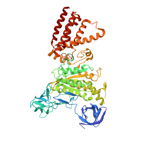
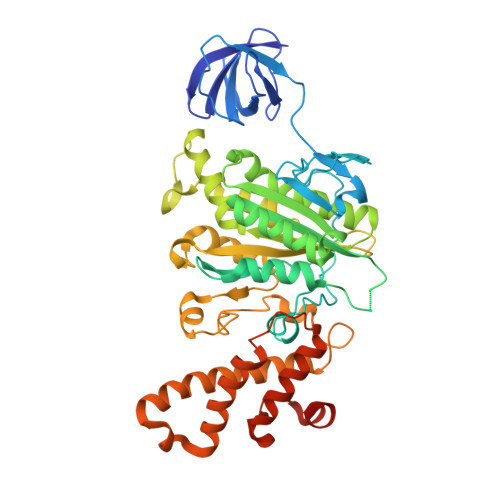
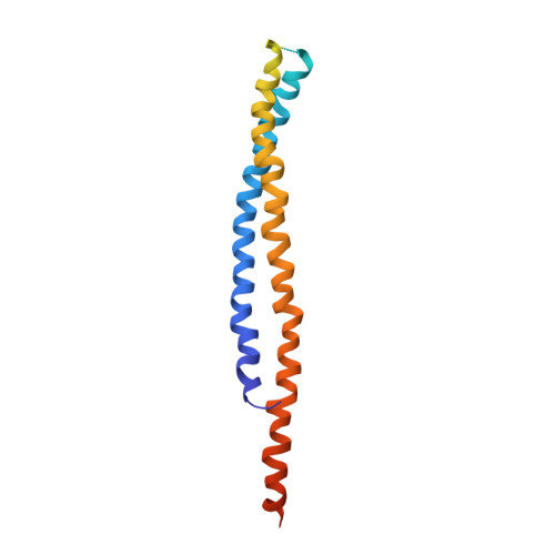
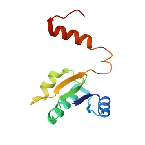
    V-type ATPases (V-ATPases) are categorized as rotary ATP synthase/ATPase complexes. The V-ATPases are distinct from F-ATPases in terms of their rotation scheme, architecture and subunit composition. However, there is no detailed structural information on V-ATPases despite the abundant biochemical and biophysical research. Here, we report a crystallographic study of V1-ATPase, from Thermus thermophilus, which is a soluble component consisting of A, B, D and F subunits. The structure at 4.5 A resolution reveals inter-subunit interactions and nucleotide binding. In particular, the structure of the central stalk composed of D and F subunits was shown to be characteristic of V1-ATPases. Small conformational changes of respective subunits and significant rearrangement of the quaternary structure observed in the three AB pairs were related to the interaction with the straight central stalk. The rotation mechanism is discussed based on a structural comparison between V1-ATPases and F1-ATPases.


  • Organizational Affiliation

    Department of Chemistry, Graduate School of Science, Kyoto University, Sakyo-ku, Kyoto 606-8502, Japan.


Macromolecules
Find similar proteins by:  (by identity cutoff)  |  3D Structure
Entity ID: 1
MoleculeChains Sequence LengthOrganismDetailsImage
V-type ATP synthase alpha chain
A, B, C, I, J
A, B, C, I, J, K
578Thermus thermophilus HB8Mutation(s): 0 
EC: 3.6.3.14
UniProt
Find proteins for Q56403 (Thermus thermophilus (strain ATCC 27634 / DSM 579 / HB8))
Explore Q56403 
Go to UniProtKB:  Q56403
Entity Groups  
Sequence Clusters30% Identity50% Identity70% Identity90% Identity95% Identity100% Identity
UniProt GroupQ56403
Sequence Annotations
Expand
  • Reference Sequence
Find similar proteins by:  (by identity cutoff)  |  3D Structure
Entity ID: 2
MoleculeChains Sequence LengthOrganismDetailsImage
V-type ATP synthase beta chain
D, E, F, L, M
D, E, F, L, M, N
478Thermus thermophilus HB8Mutation(s): 0 
EC: 3.6.3.14
UniProt
Find proteins for Q56404 (Thermus thermophilus (strain ATCC 27634 / DSM 579 / HB8))
Explore Q56404 
Go to UniProtKB:  Q56404
Entity Groups  
Sequence Clusters30% Identity50% Identity70% Identity90% Identity95% Identity100% Identity
UniProt GroupQ56404
Sequence Annotations
Expand
  • Reference Sequence
Find similar proteins by:  (by identity cutoff)  |  3D Structure
Entity ID: 3
MoleculeChains Sequence LengthOrganismDetailsImage
V-type ATP synthase subunit D
G, O
223Thermus thermophilus HB8Mutation(s): 0 
EC: 3.6.3.14
UniProt
Find proteins for O87880 (Thermus thermophilus (strain ATCC 27634 / DSM 579 / HB8))
Explore O87880 
Go to UniProtKB:  O87880
Entity Groups  
Sequence Clusters30% Identity50% Identity70% Identity90% Identity95% Identity100% Identity
UniProt GroupO87880
Sequence Annotations
Expand
  • Reference Sequence
Find similar proteins by:  (by identity cutoff)  |  3D Structure
Entity ID: 4
MoleculeChains Sequence LengthOrganismDetailsImage
V-type ATP synthase subunit F
H, P
104Thermus thermophilus HB8Mutation(s): 0 
EC: 3.6.3.14
UniProt
Find proteins for P74903 (Thermus thermophilus (strain ATCC 27634 / DSM 579 / HB8))
Explore P74903 
Go to UniProtKB:  P74903
Entity Groups  
Sequence Clusters30% Identity50% Identity70% Identity90% Identity95% Identity100% Identity
UniProt GroupP74903
Sequence Annotations
Expand
  • Reference Sequence
Experimental Data & Validation

Experimental Data

  • Method: X-RAY DIFFRACTION
  • Resolution: 4.51 Å
  • R-Value Free: 0.437 
  • R-Value Work: 0.429 
  • Space Group: P 3 2 1
Unit Cell:
Length ( Å )Angle ( ˚ )
a = 381.578α = 90
b = 381.578β = 90
c = 147.672γ = 120
Software Package:
Software NamePurpose
CNSrefinement
HKL-2000data reduction
HKL-2000data scaling

Structure Validation

View Full Validation Report


Ligand Structure Quality Assessment 


Entry History 

Deposition Data

Revision History  (Full details and data files)

  • Version 1.0: 2009-10-13
    Type: Initial release
  • Version 1.1: 2011-07-13
    Changes: Version format compliance
  • Version 1.2: 2023-11-01
    Changes: Data collection, Database references, Derived calculations, Refinement description