3A2H

Crystal structure of the rat vitamin D receptor ligand binding domain complexed with TEI-9647 and a synthetic peptide containing the NR2 box of DRIP 205


Experimental Data Snapshot

  • Method: X-RAY DIFFRACTION
  • Resolution: 2.50 Å
  • R-Value Free: 0.309 
  • R-Value Work: 0.229 
  • R-Value Observed: 0.233 

wwPDB Validation   3D Report Full Report


Ligand Structure Quality Assessment 


This is version 1.3 of the entry. See complete history


Literature

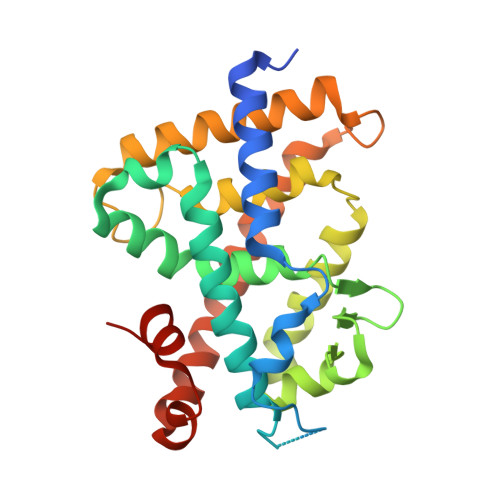
Structural basis of the histidine-mediated vitamin D receptor agonistic and antagonistic mechanisms of (23S)-25-dehydro-1alpha-hydroxyvitamin D(3)-26,23-lactone

Kakuda, S.Ishizuka, S.Eguchi, H.Mizwicki, M.T.Norman, A.W.Takimoto-Kamimura, M.

(2010) Acta Crystallogr D Biol Crystallogr 66: 918-926

  • DOI: https://doi.org/10.1107/S0907444910020810
  • Primary Citation of Related Structures:  
    3A2H, 3A2I, 3A2J

  • PubMed Abstract: 

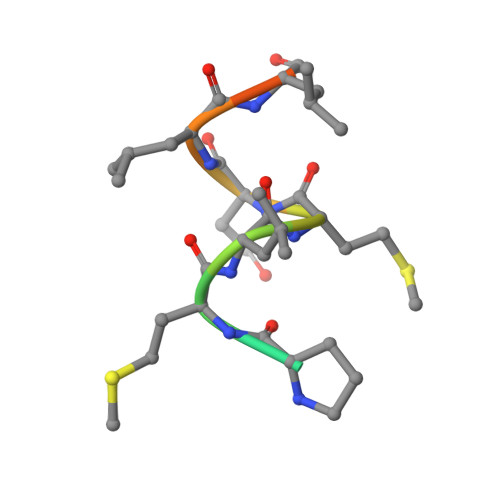
    TEI-9647 antagonizes vitamin D receptor (VDR) mediated genomic actions of 1alpha,25(OH)2D3 in human cells but is agonistic in rodent cells. The presence of Cys403, Cys410 or of both residues in the C-terminal region of human VDR (hVDR) results in antagonistic action of this compound. In the complexes of TEI-9647 with wild-type hVDR (hVDRwt) and H397F hVDR, TEI-9647 functions as an antagonist and forms a covalent adduct with hVDR according to MALDI-TOF MS. The crystal structures of complexes of TEI-9647 with rat VDR (rVDR), H305F hVDR and H305F/H397F hVDR showed that the agonistic activity of TEI-9647 is caused by a hydrogen-bond interaction with His397 or Phe397 located in helix 11. Both biological activity assays and the crystal structure of H305F hVDR complexed with TEI-9647 showed that the interaction between His305 and TEI-9647 is crucial for antagonist activity. This study indicates the following stepwise mechanism for TEI-9647 antagonism. Firstly, TEI-9647 forms hydrogen bonds to His305, which promote conformational changes in hVDR and draw Cys403 or Cys410 towards the ligand. This is followed by the formation of a 1,4-Michael addition adduct between the thiol (-SH) group of Cys403 or Cys410 and the exo-methylene group of TEI-9647.


  • Organizational Affiliation

    Teijin Institute for Bio-Medical Research, Japan.


Macromolecules
Find similar proteins by:  (by identity cutoff)  |  3D Structure
Entity ID: 1
MoleculeChains Sequence LengthOrganismDetailsImage
Vitamin D3 receptor265Rattus norvegicusMutation(s): 0 
Gene Names: VdrNr1i1
UniProt
Find proteins for P13053 (Rattus norvegicus)
Explore P13053 
Go to UniProtKB:  P13053
Entity Groups  
Sequence Clusters30% Identity50% Identity70% Identity90% Identity95% Identity100% Identity
UniProt GroupP13053
Sequence Annotations
Expand
  • Reference Sequence

Find similar proteins by:  Sequence   |   3D Structure  

Entity ID: 2
MoleculeChains Sequence LengthOrganismDetailsImage
Mediator of RNA polymerase II transcription subunit 1 peptide13Homo sapiensMutation(s): 0 
UniProt & NIH Common Fund Data Resources
Find proteins for Q15648 (Homo sapiens)
Explore Q15648 
Go to UniProtKB:  Q15648
PHAROS:  Q15648
GTEx:  ENSG00000125686 
Entity Groups  
Sequence Clusters30% Identity50% Identity70% Identity90% Identity95% Identity100% Identity
UniProt GroupQ15648
Sequence Annotations
Expand
  • Reference Sequence
Small Molecules
Ligands 1 Unique
IDChains Name / Formula / InChI Key2D Diagram3D Interactions
TEJ
Query on TEJ

Download Ideal Coordinates CCD File 
C [auth A](1S,3R,5Z,7E,20S,23S)-1,3-dihydroxy-23,26-epoxy-9,10-secocholesta-5,7,10,25(27)-tetraen-26-one
C27 H38 O4
SAODSJHDCZTVAT-HPNZEMCUSA-N
Experimental Data & Validation

Experimental Data

  • Method: X-RAY DIFFRACTION
  • Resolution: 2.50 Å
  • R-Value Free: 0.309 
  • R-Value Work: 0.229 
  • R-Value Observed: 0.233 
  • Space Group: C 1 2 1
Unit Cell:
Length ( Å )Angle ( ˚ )
a = 124.809α = 90
b = 45.349β = 100.13
c = 46.662γ = 90
Software Package:
Software NamePurpose
REFMACrefinement
CrystalCleardata collection
CrystalCleardata reduction
CrystalCleardata scaling
MOLREPphasing

Structure Validation

View Full Validation Report


Ligand Structure Quality Assessment 


Entry History 

Deposition Data

Revision History  (Full details and data files)

  • Version 1.0: 2010-05-26
    Type: Initial release
  • Version 1.1: 2011-07-13
    Changes: Version format compliance
  • Version 1.2: 2017-08-09
    Changes: Source and taxonomy
  • Version 1.3: 2023-11-01
    Changes: Data collection, Database references, Derived calculations, Refinement description