3A28

Crystal structure of L-2,3-butanediol dehydrogenase


Experimental Data Snapshot

  • Method: X-RAY DIFFRACTION
  • Resolution: 2.00 Å
  • R-Value Free: 0.240 
  • R-Value Work: 0.193 

wwPDB Validation   3D Report Full Report


Ligand Structure Quality Assessment 


This is version 1.3 of the entry. See complete history


Literature

Structural basis for chiral substrate recognition by two 2,3-butanediol dehydrogenases

Otagiri, M.Ui, S.Takusagawa, Y.Ohtsuki, T.Kurisu, G.Kusunoki, M.

(2010) FEBS Lett 584: 219-223

  • DOI: https://doi.org/10.1016/j.febslet.2009.11.068
  • Primary Citation of Related Structures:  
    3A28

  • PubMed Abstract: 

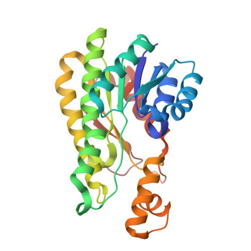
    2,3-butanediol dehydrogenase (BDH) catalyzes the NAD-dependent redox reaction between acetoin and 2,3-butanediol. There are three types of homologous BDH, each stereospecific for both substrate and product. To establish how these homologous enzymes possess differential stereospecificities, we determined the crystal structure of l-BDH with a bound inhibitor at 2.0 A. Comparison with the inhibitor binding mode of meso-BDH highlights the role of a hydrogen-bond from a conserved Trp residue(192). Site-directed mutagenesis of three active site residues of meso-BDH, including Trp(190), which corresponds to Trp(192) of L-BDH, converted its stereospecificity to that of L-BDH. This result confirms the importance of conserved residues in modifying the stereospecificity of homologous enzymes.


  • Organizational Affiliation

    Laboratory of Environmental Molecular Biology, RIKEN, Tsurumi-ward, Kanagawa, Japan.


Macromolecules
Find similar proteins by:  (by identity cutoff)  |  3D Structure
Entity ID: 1
MoleculeChains Sequence LengthOrganismDetailsImage
L-2.3-butanediol dehydrogenase258Corynebacterium glutamicumMutation(s): 0 
UniProt
Find proteins for Q9ZNN8 (Corynebacterium glutamicum)
Explore Q9ZNN8 
Go to UniProtKB:  Q9ZNN8
Entity Groups  
Sequence Clusters30% Identity50% Identity70% Identity90% Identity95% Identity100% Identity
UniProt GroupQ9ZNN8
Sequence Annotations
Expand
  • Reference Sequence
Small Molecules
Ligands 3 Unique
IDChains Name / Formula / InChI Key2D Diagram3D Interactions
NAD
Query on NAD

Download Ideal Coordinates CCD File 
AA [auth H]
I [auth C]
L [auth D]
O [auth A]
Q [auth B]
AA [auth H],
I [auth C],
L [auth D],
O [auth A],
Q [auth B],
S [auth E],
V [auth F],
Y [auth G]
NICOTINAMIDE-ADENINE-DINUCLEOTIDE
C21 H27 N7 O14 P2
BAWFJGJZGIEFAR-NNYOXOHSSA-N
BME
Query on BME

Download Ideal Coordinates CCD File 
BA [auth H]
J [auth C]
M [auth D]
P [auth A]
R [auth B]
BA [auth H],
J [auth C],
M [auth D],
P [auth A],
R [auth B],
T [auth E],
W [auth F],
Z [auth G]
BETA-MERCAPTOETHANOL
C2 H6 O S
DGVVWUTYPXICAM-UHFFFAOYSA-N
MG
Query on MG

Download Ideal Coordinates CCD File 
K [auth C],
N [auth D],
U [auth E],
X [auth F]
MAGNESIUM ION
Mg
JLVVSXFLKOJNIY-UHFFFAOYSA-N
Experimental Data & Validation

Experimental Data

  • Method: X-RAY DIFFRACTION
  • Resolution: 2.00 Å
  • R-Value Free: 0.240 
  • R-Value Work: 0.193 
  • Space Group: P 1
Unit Cell:
Length ( Å )Angle ( ˚ )
a = 60.8α = 96.1
b = 69.2β = 100.2
c = 127.4γ = 109.6
Software Package:
Software NamePurpose
ADSCdata collection
AMoREphasing
CNSrefinement
HKL-2000data reduction
HKL-2000data scaling

Structure Validation

View Full Validation Report


Ligand Structure Quality Assessment 


Entry History 

Deposition Data

Revision History  (Full details and data files)

  • Version 1.0: 2009-12-15
    Type: Initial release
  • Version 1.1: 2011-07-13
    Changes: Version format compliance
  • Version 1.2: 2014-01-22
    Changes: Database references
  • Version 1.3: 2023-11-01
    Changes: Data collection, Database references, Derived calculations, Refinement description