


3A23
Crystal Structure of beta-L-Arabinopyranosidase complexed with D-galactose
- PDB DOI: https://doi.org/10.2210/pdb3A23/pdb
- Classification: HYDROLASE
- Organism(s): Streptomyces avermitilis
- Expression System: Streptomyces lividans
- Mutation(s): No
- Deposited: 2009-04-27 Released: 2009-07-14
Experimental Data Snapshot
- Method: X-RAY DIFFRACTION
- Resolution: 1.90 Å
- R-Value Free: 0.177
- R-Value Work: 0.142
- R-Value Observed: 0.144

This is version 1.3 of the entry. See complete history.

Macromolecules
Find similar proteins by:
(by identity cutoff) | 3D Structure
Entity ID: 1 | |||||
|---|---|---|---|---|---|
| Molecule | Chains | Sequence Length | Organism | Details | Image |
| Putative secreted alpha-galactosidase | 614 | Streptomyces avermitilis | Mutation(s): 0 Gene Names: agaA2, SAV2186, SAV_2186 | 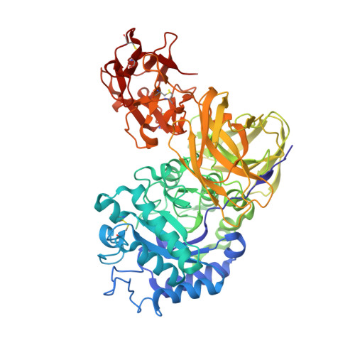 | |
UniProt | |||||
Find proteins for Q82L26 (Streptomyces avermitilis (strain ATCC 31267 / DSM 46492 / JCM 5070 / NBRC 14893 / NCIMB 12804 / NRRL 8165 / MA-4680)) Explore Q82L26 Go to UniProtKB: Q82L26 | |||||
Entity Groups | |||||
| Sequence Clusters | 30% Identity50% Identity70% Identity90% Identity95% Identity100% Identity | ||||
| UniProt Group | Q82L26 | ||||
Sequence AnnotationsExpand | |||||
| |||||
Small Molecules
| Ligands 5 Unique | |||||
|---|---|---|---|---|---|
| ID | Chains | Name / Formula / InChI Key | 2D Diagram | 3D Interactions | |
| 1PG Query on 1PG | DA [auth B], P [auth A] | 2-(2-{2-[2-(2-METHOXY-ETHOXY)-ETHOXY]-ETHOXY}-ETHOXY)-ETHANOL C11 H24 O6 SLNYBUIEAMRFSZ-UHFFFAOYSA-N |  | ||
| EPE Query on EPE | CA [auth B] | 4-(2-HYDROXYETHYL)-1-PIPERAZINE ETHANESULFONIC ACID C8 H18 N2 O4 S JKMHFZQWWAIEOD-UHFFFAOYSA-N |  | ||
| GAL Query on GAL | C [auth A], T [auth B], U [auth B] | beta-D-galactopyranose C6 H12 O6 WQZGKKKJIJFFOK-FPRJBGLDSA-N |  | ||
| SO4 Query on SO4 | EA [auth B], FA [auth B], Q [auth A], R [auth A], S [auth A] | SULFATE ION O4 S QAOWNCQODCNURD-UHFFFAOYSA-L |  | ||
| GOL Query on GOL | AA [auth B] BA [auth B] D [auth A] E [auth A] F [auth A] | GLYCEROL C3 H8 O3 PEDCQBHIVMGVHV-UHFFFAOYSA-N |  | ||
Experimental Data & Validation
Experimental Data
- Method: X-RAY DIFFRACTION
- Resolution: 1.90 Å
- R-Value Free: 0.177
- R-Value Work: 0.142
- R-Value Observed: 0.144
- Space Group: P 21 21 21
Unit Cell:
| Length ( Å ) | Angle ( ˚ ) |
|---|---|
| a = 68.256 | α = 90 |
| b = 99.081 | β = 90 |
| c = 181.63 | γ = 90 |
| Software Name | Purpose |
|---|---|
| REFMAC | refinement |
| HKL-2000 | data collection |
| HKL-2000 | data reduction |
| HKL-2000 | data scaling |
| REFMAC | phasing |
Entry History
Deposition Data
- Released Date: 2009-07-14 Deposition Author(s): Fujimoto, Z., Ichinose, H., Kaneko, S.
Revision History (Full details and data files)
- Version 1.0: 2009-07-14
Type: Initial release - Version 1.1: 2011-07-13
Changes: Non-polymer description, Version format compliance - Version 1.2: 2020-07-29
Type: Remediation
Reason: Carbohydrate remediation
Changes: Data collection, Derived calculations, Structure summary - Version 1.3: 2023-11-01
Changes: Data collection, Database references, Refinement description, Structure summary
