3A1J

Crystal structure of the human Rad9-Hus1-Rad1 complex


Experimental Data Snapshot

  • Method: X-RAY DIFFRACTION
  • Resolution: 2.50 Å
  • R-Value Free: 0.282 
  • R-Value Work: 0.209 
  • R-Value Observed: 0.209 

wwPDB Validation   3D Report Full Report


This is version 2.0 of the entry. See complete history


Literature

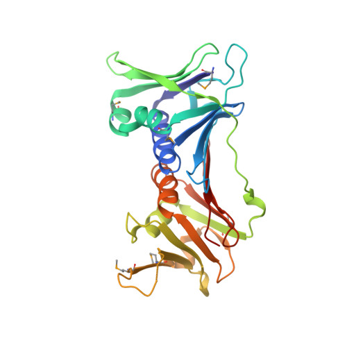
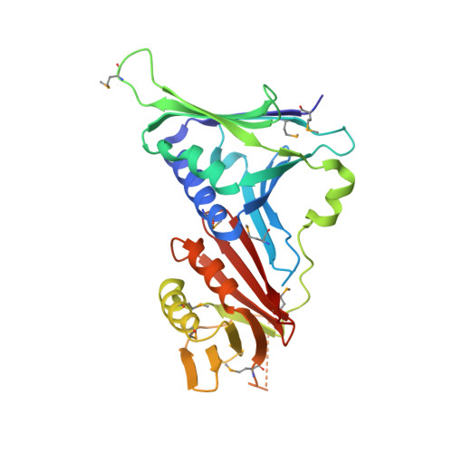
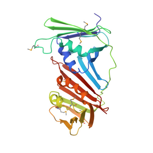
Crystal Structure of the Human Rad9-Hus1-Rad1 Clamp

Sohn, S.Y.Cho, Y.

(2009) J Mol Biol 390: 490-502

  • DOI: https://doi.org/10.1016/j.jmb.2009.05.028
  • Primary Citation of Related Structures:  
    3A1J

  • PubMed Abstract: 

    Three evolutionarily conserved proteins, Rad9, Hus1, and Rad1, form a heterotrimeric 9-1-1 complex that plays critical roles in cellular responses to DNA damage by activating checkpoints and by recruiting DNA repair enzymes to DNA lesions. We have determined the crystal structure of the human Rad9 (residues 1-272)-Hus1-Rad1 complex at 2.5 A resolution. The 9(1-272)-1-1 complex forms a closed ring, with each subunit having a similar structure. Despite its high level of similarity to proliferating cell nucleus antigen in terms of overall structure, the 9(1-272)-1-1 complex exhibits notable differences in local structures, including interdomain connecting loops, H2 and H3 helices, and loops in the vicinity of the helices of each subunit. These local structural variations provide several unique features to the 9-1-1 heterotrimeric complex-including structures of intermolecular interfaces and the inner surface around the central hole, and different electrostatic potentials at and near the interdomain connecting loops of each 9-1-1 subunit-compared to the proliferating cell nucleus antigen trimer. We propose that these structural features allow the 9-1-1 complex to bind to a damaged DNA during checkpoint control and to serve as a platform for base excision repair. We also show that the 9(1-272)-1-1 complex, but not the full-length 9-1-1 complex, forms a stable complex with the 5' recessed DNA, suggesting that the C-terminal tail of Rad9 is involved in the regulation of the 9-1-1 complex in DNA binding.


  • Organizational Affiliation

    National Creative Research Center for Structural Biology and Department of Life Science, Pohang University of Science and Technology, Hyo-ja dong, Pohang, KyungBook, South Korea.


Macromolecules
Find similar proteins by:  (by identity cutoff)  |  3D Structure
Entity ID: 1
MoleculeChains Sequence LengthOrganismDetailsImage
Cell cycle checkpoint control protein RAD9A266Homo sapiensMutation(s): 0 
Gene Names: Rad9(1-272)
EC: 3.1.11.2
UniProt & NIH Common Fund Data Resources
Find proteins for Q99638 (Homo sapiens)
Explore Q99638 
Go to UniProtKB:  Q99638
PHAROS:  Q99638
GTEx:  ENSG00000172613 
Entity Groups  
Sequence Clusters30% Identity50% Identity70% Identity90% Identity95% Identity100% Identity
UniProt GroupQ99638
Sequence Annotations
Expand
  • Reference Sequence
Find similar proteins by:  (by identity cutoff)  |  3D Structure
Entity ID: 2
MoleculeChains Sequence LengthOrganismDetailsImage
Checkpoint protein HUS1281Homo sapiensMutation(s): 0 
Gene Names: HUS1
UniProt & NIH Common Fund Data Resources
Find proteins for O60921 (Homo sapiens)
Explore O60921 
Go to UniProtKB:  O60921
PHAROS:  O60921
GTEx:  ENSG00000136273 
Entity Groups  
Sequence Clusters30% Identity50% Identity70% Identity90% Identity95% Identity100% Identity
UniProt GroupO60921
Sequence Annotations
Expand
  • Reference Sequence
Find similar proteins by:  (by identity cutoff)  |  3D Structure
Entity ID: 3
MoleculeChains Sequence LengthOrganismDetailsImage
Cell cycle checkpoint protein RAD1263Homo sapiensMutation(s): 0 
Gene Names: RAD1
EC: 3.1.11.2
UniProt & NIH Common Fund Data Resources
Find proteins for O60671 (Homo sapiens)
Explore O60671 
Go to UniProtKB:  O60671
PHAROS:  O60671
GTEx:  ENSG00000113456 
Entity Groups  
Sequence Clusters30% Identity50% Identity70% Identity90% Identity95% Identity100% Identity
UniProt GroupO60671
Sequence Annotations
Expand
  • Reference Sequence
Oligosaccharides

Help

Entity ID: 4
MoleculeChains Length2D Diagram Glycosylation3D Interactions
beta-D-fructofuranose-(2-1)-alpha-D-glucopyranose
D
2N/A
Glycosylation Resources
GlyTouCan:  G05551OP
GlyCosmos:  G05551OP
Small Molecules
Modified Residues  1 Unique
IDChains TypeFormula2D DiagramParent
MSE
Query on MSE
A
L-PEPTIDE LINKINGC5 H11 N O2 SeMET
Biologically Interesting Molecules (External Reference) 1 Unique
Experimental Data & Validation

Experimental Data

  • Method: X-RAY DIFFRACTION
  • Resolution: 2.50 Å
  • R-Value Free: 0.282 
  • R-Value Work: 0.209 
  • R-Value Observed: 0.209 
  • Space Group: P 1 21 1
Unit Cell:
Length ( Å )Angle ( ˚ )
a = 77.154α = 90
b = 70.776β = 99.2
c = 86.661γ = 90
Software Package:
Software NamePurpose
CNSrefinement
HKL-2000data collection
HKL-2000data reduction
HKL-2000data scaling
SOLVEphasing

Structure Validation

View Full Validation Report


Entry History 

Deposition Data

Revision History  (Full details and data files)

  • Version 1.0: 2009-06-02
    Type: Initial release
  • Version 1.1: 2011-07-13
    Changes: Version format compliance
  • Version 2.0: 2020-07-29
    Type: Remediation
    Reason: Carbohydrate remediation
    Changes: Atomic model, Data collection, Database references, Derived calculations, Non-polymer description, Structure summary