


3WJ1
Crystal structure of SSHESTI
- PDB DOI: https://doi.org/10.2210/pdb3WJ1/pdb
- Classification: HYDROLASE
- Organism(s): Saccharolobus shibatae
- Expression System: Escherichia coli
- Mutation(s): No
- Deposited: 2013-10-03 Released: 2014-07-30
Experimental Data Snapshot
- Method: X-RAY DIFFRACTION
- Resolution: 1.50 Å
- R-Value Free: 0.180
- R-Value Work: 0.157
- R-Value Observed: 0.158

This is version 1.2 of the entry. See complete history.

Macromolecules
Find similar proteins by:
(by identity cutoff) | 3D Structure
Entity ID: 1 | |||||
|---|---|---|---|---|---|
| Molecule | Chains | Sequence Length | Organism | Details | Image |
| Carboxylesterase | 305 | Saccharolobus shibatae | Mutation(s): 0 Gene Names: SshEstI EC: 3.1.1.1 | 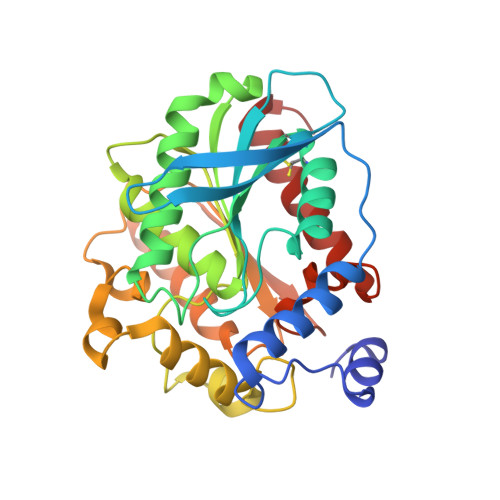 | |
UniProt | |||||
Find proteins for Q5NU42 (Saccharolobus shibatae) Explore Q5NU42 Go to UniProtKB: Q5NU42 | |||||
Entity Groups | |||||
| Sequence Clusters | 30% Identity50% Identity70% Identity90% Identity95% Identity100% Identity | ||||
| UniProt Group | Q5NU42 | ||||
Sequence AnnotationsExpand | |||||
| |||||
Small Molecules
| Ligands 1 Unique | |||||
|---|---|---|---|---|---|
| ID | Chains | Name / Formula / InChI Key | 2D Diagram | 3D Interactions | |
| BOG Query on BOG | B [auth A] | octyl beta-D-glucopyranoside C14 H28 O6 HEGSGKPQLMEBJL-RKQHYHRCSA-N |  | ||
Experimental Data & Validation
Experimental Data
- Method: X-RAY DIFFRACTION
- Resolution: 1.50 Å
- R-Value Free: 0.180
- R-Value Work: 0.157
- R-Value Observed: 0.158
- Space Group: I 2 2 2
Unit Cell:
| Length ( Å ) | Angle ( ˚ ) |
|---|---|
| a = 58.403 | α = 90 |
| b = 71.942 | β = 90 |
| c = 137.332 | γ = 90 |
| Software Name | Purpose |
|---|---|
| MOLREP | phasing |
| REFMAC | refinement |
| MOSFLM | data reduction |
| SCALA | data scaling |
Entry History
Deposition Data
- Released Date: 2014-07-30 Deposition Author(s): Ohara, K., Unno, H., Oshima, Y., Furukawa, K., Fujino, N., Hirooka, K., Hemmi, H., Takahashi, S., Nishino, T., Kusunoki, M., Nakayama, T.
Revision History (Full details and data files)
- Version 1.0: 2014-07-30
Type: Initial release - Version 1.1: 2019-12-25
Changes: Database references, Derived calculations - Version 1.2: 2020-07-29
Type: Remediation
Reason: Carbohydrate remediation
Changes: Data collection, Derived calculations, Structure summary
