3WIH

Crystal structure of the third fibronectin domain (Fn3) of human ROBO1 in complex with the Fab fragment of murine monoclonal antibody B2212A.


Experimental Data Snapshot

  • Method: X-RAY DIFFRACTION
  • Resolution: 1.70 Å
  • R-Value Free: 0.236 
  • R-Value Work: 0.189 
  • R-Value Observed: 0.192 

wwPDB Validation   3D Report Full Report


This is version 1.3 of the entry. See complete history


Literature

Structural features of interfacial tyrosine residue in ROBO1 fibronectin domain-antibody complex: Crystallographic, thermodynamic, and molecular dynamic analyses

Nakayama, T.Mizohata, E.Yamashita, T.Nagatoishi, S.Nakakido, M.Iwanari, H.Mochizuki, Y.Kado, Y.Yokota, Y.Satoh, R.Tsumoto, K.Fujitani, H.Kodama, T.Hamakubo, T.Inoue, T.

(2015) Protein Sci 24: 328-340

  • DOI: https://doi.org/10.1002/pro.2619
  • Primary Citation of Related Structures:  
    3WIH, 3WII

  • PubMed Abstract: 

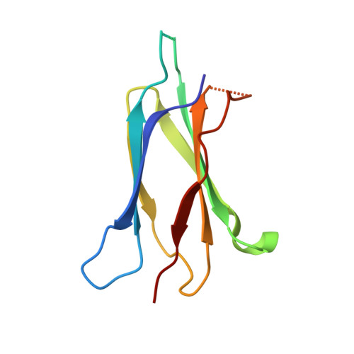
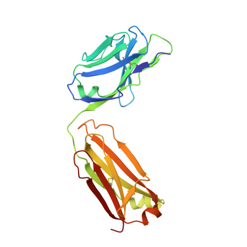
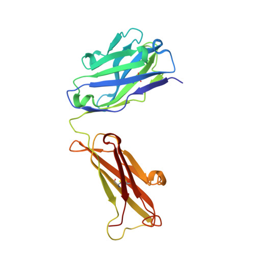
    ROBO1, fibronectin Type-III domain (Fn)-containing protein, is a novel immunotherapeutic target for hepatocellular carcinoma in humans. The crystal structure of the antigen-binding fragment (Fab) of B2212A, the monoclonal antibody against the third Fn domain (Fn3) of ROBO1, was determined in pursuit of antibody drug for hepatocellular carcinoma. This effort was conducted in the presence or absence of the antigen, with the chemical features being investigated by determining the affinity of the antibody using molecular dynamics (MD) and thermodynamics. The structural comparison of B2212A Fab between the complex and the free form revealed that the interfacial Tyr(L) 50 (superscripts L, H, and F stand for the residues in the light chain, heavy chain, and Fn3, respectively) played important roles in Fn3 recognition. That is, the aromatic ring of Tyr(L) 50 pivoted toward Phe(F) 68, forming a CH/π interaction and a new hydrogen bond with the carbonyl O atom of Phe(F) 68. MD simulations predicted that the Tyr(L) 50-Phe(F) 68 interaction almost entirely dominated Fab-Fn3 binding, and Ala-substitution of Tyr(L) 50 led to a reduced binding of the resultant complex. On the contrary, isothermal titration calorimetry experiments underscored that Ala-substitution of Tyr(L) 50 caused an increase of the binding enthalpy between B2212A and Fn3, but importantly, it induced an increase of the binding entropy, resulting in a suppression of loss in the Gibbs free energy in total. These results suggest that mutation analysis considering the binding entropy as well as the binding enthalpy will aid in the development of novel antibody drugs for hepatocellular carcinoma.


  • Organizational Affiliation

    Structural Physical Chemistry, Division of Applied Chemistry, Graduate School of Engineering, Osaka University, Osaka, Japan.


Macromolecules
Find similar proteins by:  (by identity cutoff)  |  3D Structure
Entity ID: 1
MoleculeChains Sequence LengthOrganismDetailsImage
Roundabout homolog 1A,
D [auth B]
97Homo sapiensMutation(s): 0 
Gene Names: DUTT1ROBO1Roundabout homolog 1
UniProt & NIH Common Fund Data Resources
Find proteins for Q9Y6N7 (Homo sapiens)
Explore Q9Y6N7 
Go to UniProtKB:  Q9Y6N7
PHAROS:  Q9Y6N7
GTEx:  ENSG00000169855 
Entity Groups  
Sequence Clusters30% Identity50% Identity70% Identity90% Identity95% Identity100% Identity
UniProt GroupQ9Y6N7
Sequence Annotations
Expand
  • Reference Sequence
Find similar proteins by:  (by identity cutoff)  |  3D Structure
Entity ID: 2
MoleculeChains Sequence LengthOrganismDetailsImage
anti-human ROBO1 antibody B2212A Fab light chainB [auth L],
E [auth M]
213Mus musculusMutation(s): 0 
Entity Groups  
Sequence Clusters30% Identity50% Identity70% Identity90% Identity95% Identity100% Identity
Sequence Annotations
Expand
  • Reference Sequence
Find similar proteins by:  (by identity cutoff)  |  3D Structure
Entity ID: 3
MoleculeChains Sequence LengthOrganismDetailsImage
anti-human ROBO1 antibody B2212A Fab heavy chainC [auth H],
F [auth I]
219Mus musculusMutation(s): 0 
Entity Groups  
Sequence Clusters30% Identity50% Identity70% Identity90% Identity95% Identity100% Identity
Sequence Annotations
Expand
  • Reference Sequence
Experimental Data & Validation

Experimental Data

  • Method: X-RAY DIFFRACTION
  • Resolution: 1.70 Å
  • R-Value Free: 0.236 
  • R-Value Work: 0.189 
  • R-Value Observed: 0.192 
  • Space Group: C 1 2 1
Unit Cell:
Length ( Å )Angle ( ˚ )
a = 159.474α = 90
b = 102.601β = 127.57
c = 97.236γ = 90
Software Package:
Software NamePurpose
DENZOdata reduction
SCALEPACKdata scaling
REFMACrefinement
PDB_EXTRACTdata extraction
BSSdata collection
HKL-2000data reduction
HKL-2000data scaling
MOLREPphasing
PHENIXrefinement

Structure Validation

View Full Validation Report


Entry History 

Revision History  (Full details and data files)

  • Version 1.0: 2015-01-21
    Type: Initial release
  • Version 1.1: 2015-03-04
    Changes: Database references
  • Version 1.2: 2017-11-22
    Changes: Refinement description
  • Version 1.3: 2023-11-08
    Changes: Data collection, Database references, Derived calculations, Refinement description