3WBD

Crystal structure of anti-polysialic acid antibody single chain Fv fragment (mAb735) complexed with octasialic acid


Experimental Data Snapshot

  • Method: X-RAY DIFFRACTION
  • Resolution: 1.80 Å
  • R-Value Free: 0.259 
  • R-Value Work: 0.233 
  • R-Value Observed: 0.234 

wwPDB Validation   3D Report Full Report


This is version 2.1 of the entry. See complete history


Literature

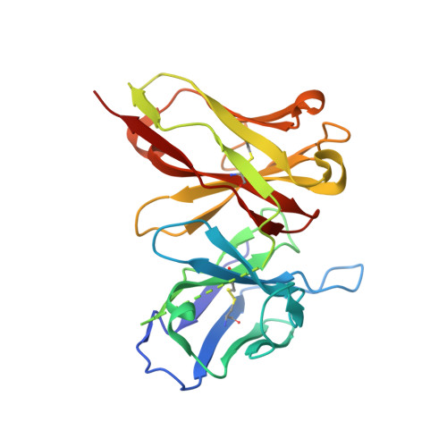
Crystal structure of anti-polysialic acid antibody single chain Fv fragment complexed with octasialic acid: insight into the binding preference for polysialic acid.

Nagae, M.Ikeda, A.Hane, M.Hanashima, S.Kitajima, K.Sato, C.Yamaguchi, Y.

(2013) J Biol Chem 288: 33784-33796

  • DOI: https://doi.org/10.1074/jbc.M113.496224
  • Primary Citation of Related Structures:  
    3WBD

  • PubMed Abstract: 

    Polysialic acid is a linear homopolymer of α2-8-linked sialic acids attached mainly onto glycoproteins. Cell surface polysialic acid plays roles in cell adhesion and differentiation events in a manner that is often dependent on the degree of polymerization (DP). Anti-oligo/polysialic acid antibodies have DP-dependent antigenic specificity, and such antibodies are widely utilized in biological studies for detecting and distinguishing between different oligo/polysialic acids. A murine monoclonal antibody mAb735 has a unique preference for longer polymers of polysialic acid (DP >10), yet the mechanism of recognition at the atomic level remains unclear. Here, we report the crystal structure of mAb735 single chain variable fragment (scFv735) in complex with octasialic acid at 1.8 Å resolution. In the asymmetric unit, two scFv735 molecules associate with one octasialic acid. In both complexes of the unit, all the complementarity-determining regions except for L3 interact with three consecutive sialic acid residues out of the eight. A striking feature of the complex is that 11 ordered water molecules bridge the gap between antibody and ligand, whereas the direct antibody-ligand interaction is less extensive. The dihedral angles of the trisialic acid unit directly interacting with scFv735 are not uniform, indicating that mAb735 does not strictly favor the previously proposed helical conformation. Importantly, both reducing and nonreducing ends of the bound ligand are completely exposed to solvent. We suggest that mAb735 gains its apparent high affinity for a longer polysialic acid chain by recognizing every three sialic acid units in a paired manner.


  • Organizational Affiliation

    Structural Glycobiology Team, Systems Glycobiology Research Group, RIKEN-Max Planck Joint Research Center, RIKEN Global Research Cluster, 2-1 Hirosawa, Wako, Saitama 351-0198.


Macromolecules
Find similar proteins by:  (by identity cutoff)  |  3D Structure
Entity ID: 1
MoleculeChains Sequence LengthOrganismDetailsImage
single chain Fv fragment of mAb735
A, B
246Mus musculusMutation(s): 0 
Entity Groups  
Sequence Clusters30% Identity50% Identity70% Identity90% Identity95% Identity100% Identity
Sequence Annotations
Expand
  • Reference Sequence
Oligosaccharides

Help

Entity ID: 2
MoleculeChains Length2D Diagram Glycosylation3D Interactions
N-acetyl-alpha-neuraminic acid-(2-8)-N-acetyl-alpha-neuraminic acid-(2-8)-N-acetyl-alpha-neuraminic acid-(2-8)-N-acetyl-alpha-neuraminic acid-(2-8)-N-acetyl-alpha-neuraminic acid-(2-8)-N-acetyl-alpha-neuraminic acid-(2-8)-N-acetyl-alpha-neuraminic acid
C
7N/A
Glycosylation Resources
GlyTouCan:  G70323IQ
GlyCosmos:  G70323IQ
Small Molecules
Ligands 1 Unique
IDChains Name / Formula / InChI Key2D Diagram3D Interactions
FLC
Query on FLC

Download Ideal Coordinates CCD File 
D [auth A]CITRATE ANION
C6 H5 O7
KRKNYBCHXYNGOX-UHFFFAOYSA-K
Experimental Data & Validation

Experimental Data

  • Method: X-RAY DIFFRACTION
  • Resolution: 1.80 Å
  • R-Value Free: 0.259 
  • R-Value Work: 0.233 
  • R-Value Observed: 0.234 
  • Space Group: P 64
Unit Cell:
Length ( Å )Angle ( ˚ )
a = 161.174α = 90
b = 161.174β = 90
c = 45.884γ = 120
Software Package:
Software NamePurpose
SERGUIdata collection
MOLREPphasing
REFMACrefinement
HKL-2000data reduction
HKL-2000data scaling

Structure Validation

View Full Validation Report


Entry History 

Deposition Data

Revision History  (Full details and data files)

  • Version 1.0: 2013-10-16
    Type: Initial release
  • Version 1.1: 2014-04-02
    Changes: Database references
  • Version 1.2: 2014-04-16
    Changes: Source and taxonomy
  • Version 1.3: 2017-11-22
    Changes: Refinement description
  • Version 2.0: 2020-07-29
    Type: Remediation
    Reason: Carbohydrate remediation
    Changes: Atomic model, Data collection, Derived calculations, Structure summary
  • Version 2.1: 2023-11-08
    Changes: Data collection, Database references, Refinement description, Structure summary