3W1V

Crystal Structure of Capsular Polysaccharide Synthesizing Enzyme CapE from Staphylococcus aureus in complex with inihibitor


Experimental Data Snapshot

  • Method: X-RAY DIFFRACTION
  • Resolution: 2.10 Å
  • R-Value Free: 0.207 
  • R-Value Work: 0.164 
  • R-Value Observed: 0.166 

wwPDB Validation   3D Report Full Report


Ligand Structure Quality Assessment 


This is version 1.3 of the entry. See complete history


Literature

Crystal structure of the capsular polysaccharide synthesizing protein CapE of Staphylococcus aureus.

Miyafusa, T.Caaveiro, J.M.Tanaka, Y.Tanner, M.E.Tsumoto, K.

(2013) Biosci Rep 33: 463-474

  • DOI: https://doi.org/10.1042/BSR20130017
  • Primary Citation of Related Structures:  
    3VVB, 3VVC, 3W1V

  • PubMed Abstract: 

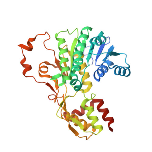
    Enzymes synthesizing the bacterial CP (capsular polysaccharide) are attractive antimicrobial targets. However, we lack critical information about the structure and mechanism of many of them. In an effort to reduce that gap, we have determined three different crystal structures of the enzyme CapE of the human pathogen Staphylococcus aureus. The structure reveals that CapE is a member of the SDR (short-chain dehydrogenase/reductase) super-family of proteins. CapE assembles in a hexameric complex stabilized by three major contact surfaces between protein subunits. Turnover of substrate and/or coenzyme induces major conformational changes at the contact interface between protein subunits, and a displacement of the substrate-binding domain with respect to the Rossmann domain. A novel dynamic element that we called the latch is essential for remodelling of the protein-protein interface. Structural and primary sequence alignment identifies a group of SDR proteins involved in polysaccharide synthesis that share the two salient features of CapE: the mobile loop (latch) and a distinctive catalytic site (MxxxK). The relevance of these structural elements was evaluated by site-directed mutagenesis.


  • Organizational Affiliation

    Medical Proteomics Laboratory, Institute of Medical Science, Department of Medical Genome Sciences, School of Frontier Sciences, The University of Tokyo, Minato-ku, Tokyo 108-8639, Japan.


Macromolecules
Find similar proteins by:  (by identity cutoff)  |  3D Structure
Entity ID: 1
MoleculeChains Sequence LengthOrganismDetailsImage
Capsular polysaccharide synthesis enzyme Cap8E
A, B
363Staphylococcus aureus subsp. aureus Mu50Mutation(s): 0 
Gene Names: capE
EC: 4.2.1.115
UniProt
Find proteins for A0A0H3JPH0 (Staphylococcus aureus (strain Mu50 / ATCC 700699))
Explore A0A0H3JPH0 
Go to UniProtKB:  A0A0H3JPH0
Entity Groups  
Sequence Clusters30% Identity50% Identity70% Identity90% Identity95% Identity100% Identity
UniProt GroupA0A0H3JPH0
Sequence Annotations
Expand
  • Reference Sequence
Small Molecules
Ligands 4 Unique
IDChains Name / Formula / InChI Key2D Diagram3D Interactions
UDZ
Query on UDZ

Download Ideal Coordinates CCD File 
K [auth A],
T [auth B]
[(2R,3S,4R,5R,6R)-5-acetamido-6-[[[(2R,3S,4R,5R)-5-[2,4-bis(oxidanylidene)pyrimidin-1-yl]-3,4-bis(oxidanyl)oxolan-2-yl]methoxy-oxidanyl-phosphoryl]oxy-oxidanyl-phosphoryl]oxy-3,4-bis(oxidanyl)oxan-2-yl]methylimino-azanylidene-azanium
C17 H27 N6 O16 P2
JRORMXITLCSMOP-CFRASDGPSA-O
EPE
Query on EPE

Download Ideal Coordinates CCD File 
J [auth A]4-(2-HYDROXYETHYL)-1-PIPERAZINE ETHANESULFONIC ACID
C8 H18 N2 O4 S
JKMHFZQWWAIEOD-UHFFFAOYSA-N
SO4
Query on SO4

Download Ideal Coordinates CCD File 
C [auth A]
D [auth A]
E [auth A]
F [auth A]
G [auth A]
C [auth A],
D [auth A],
E [auth A],
F [auth A],
G [auth A],
H [auth A],
I [auth A],
L [auth B],
M [auth B],
N [auth B],
O [auth B],
P [auth B],
Q [auth B],
R [auth B]
SULFATE ION
O4 S
QAOWNCQODCNURD-UHFFFAOYSA-L
NA
Query on NA

Download Ideal Coordinates CCD File 
S [auth B]SODIUM ION
Na
FKNQFGJONOIPTF-UHFFFAOYSA-N
Experimental Data & Validation

Experimental Data

  • Method: X-RAY DIFFRACTION
  • Resolution: 2.10 Å
  • R-Value Free: 0.207 
  • R-Value Work: 0.164 
  • R-Value Observed: 0.166 
  • Space Group: P 3 2 1
Unit Cell:
Length ( Å )Angle ( ˚ )
a = 123.78α = 90
b = 123.78β = 90
c = 104.46γ = 120
Software Package:
Software NamePurpose
SERGUIdata collection
PHASERphasing
REFMACrefinement
MOSFLMdata reduction
SCALAdata scaling

Structure Validation

View Full Validation Report


Ligand Structure Quality Assessment 


Entry History 

Deposition Data

Revision History  (Full details and data files)

  • Version 1.0: 2013-06-12
    Type: Initial release
  • Version 1.1: 2014-01-29
    Changes: Database references
  • Version 1.2: 2017-11-22
    Changes: Refinement description
  • Version 1.3: 2024-03-20
    Changes: Data collection, Database references, Derived calculations