


3VM6
Crystal structure of ribose-1,5-bisphosphate isomerase from Thermococcus kodakarensis KOD1 in complex with alpha-D-ribose-1,5-bisphosphate
- PDB DOI: https://doi.org/10.2210/pdb3VM6/pdb
- Classification: ISOMERASE
- Organism(s): Thermococcus kodakarensis KOD1
- Expression System: Escherichia coli
- Mutation(s): Yes
- Deposited: 2011-12-08 Released: 2012-04-25
Experimental Data Snapshot
- Method: X-RAY DIFFRACTION
- Resolution: 2.85 Å
- R-Value Free: 0.262
- R-Value Work: 0.201
- R-Value Observed: 0.201

This is version 1.3 of the entry. See complete history.

Macromolecules
Find similar proteins by:
(by identity cutoff) | 3D Structure
Entity ID: 1 | |||||
|---|---|---|---|---|---|
| Molecule | Chains | Sequence Length | Organism | Details | Image |
| Translation initiation factor eIF-2B, delta subunit | 338 | Thermococcus kodakarensis KOD1 | Mutation(s): 1 Gene Names: E2b2, TK0185 EC: 5.3.1 | 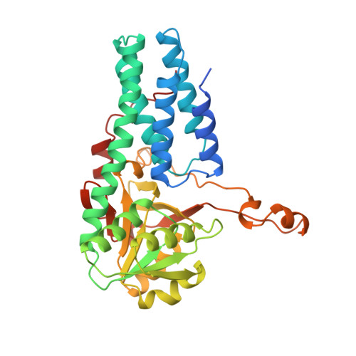 | |
UniProt | |||||
Find proteins for Q5JFM9 (Thermococcus kodakarensis (strain ATCC BAA-918 / JCM 12380 / KOD1)) Explore Q5JFM9 Go to UniProtKB: Q5JFM9 | |||||
Entity Groups | |||||
| Sequence Clusters | 30% Identity50% Identity70% Identity90% Identity95% Identity100% Identity | ||||
| UniProt Group | Q5JFM9 | ||||
Sequence AnnotationsExpand | |||||
| |||||
Small Molecules
| Ligands 5 Unique | |||||
|---|---|---|---|---|---|
| ID | Chains | Name / Formula / InChI Key | 2D Diagram | 3D Interactions | |
| RI2 Query on RI2 | D [auth A], J [auth B], P [auth C] | 1,5-di-O-phosphono-alpha-D-ribofuranose C5 H12 O11 P2 AAAFZMYJJHWUPN-TXICZTDVSA-N |  | ||
| PG4 Query on PG4 | L [auth B] | TETRAETHYLENE GLYCOL C8 H18 O5 UWHCKJMYHZGTIT-UHFFFAOYSA-N |  | ||
| PEG Query on PEG | E [auth A] F [auth A] G [auth A] K [auth B] M [auth B] | DI(HYDROXYETHYL)ETHER C4 H10 O3 MTHSVFCYNBDYFN-UHFFFAOYSA-N |  | ||
| CL Query on CL | H [auth A], O [auth B], U [auth C] | CHLORIDE ION Cl VEXZGXHMUGYJMC-UHFFFAOYSA-M |  | ||
| MG Query on MG | I [auth A], V [auth C] | MAGNESIUM ION Mg JLVVSXFLKOJNIY-UHFFFAOYSA-N |  | ||
Experimental Data & Validation
Experimental Data
- Method: X-RAY DIFFRACTION
- Resolution: 2.85 Å
- R-Value Free: 0.262
- R-Value Work: 0.201
- R-Value Observed: 0.201
- Space Group: P 41 21 2
Unit Cell:
| Length ( Å ) | Angle ( ˚ ) |
|---|---|
| a = 146.537 | α = 90 |
| b = 146.537 | β = 90 |
| c = 99.649 | γ = 90 |
| Software Name | Purpose |
|---|---|
| HKL-2000 | data collection |
| MOLREP | phasing |
| CNS | refinement |
| HKL-2000 | data reduction |
| SCALEPACK | data scaling |
Entry History
Deposition Data
- Released Date: 2012-04-25 Deposition Author(s): Nakamura, A., Fujihashi, M., Aono, R., Sato, T., Nishiba, Y., Yoshida, S., Yano, A., Atomi, H., Imanaka, T., Miki, K.
Revision History (Full details and data files)
- Version 1.0: 2012-04-25
Type: Initial release - Version 1.1: 2013-06-12
Changes: Database references - Version 1.2: 2020-07-29
Type: Remediation
Reason: Carbohydrate remediation
Changes: Data collection, Database references, Derived calculations - Version 1.3: 2023-11-08
Changes: Data collection, Database references, Refinement description, Structure summary
