3VJ6

Structure of the MHC class Ib molecule Qa-1b


Experimental Data Snapshot

  • Method: X-RAY DIFFRACTION
  • Resolution: 1.90 Å
  • R-Value Free: 0.242 
  • R-Value Work: 0.201 
  • R-Value Observed: 0.203 

wwPDB Validation   3D Report Full Report


This is version 1.1 of the entry. See complete history


Literature

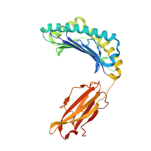
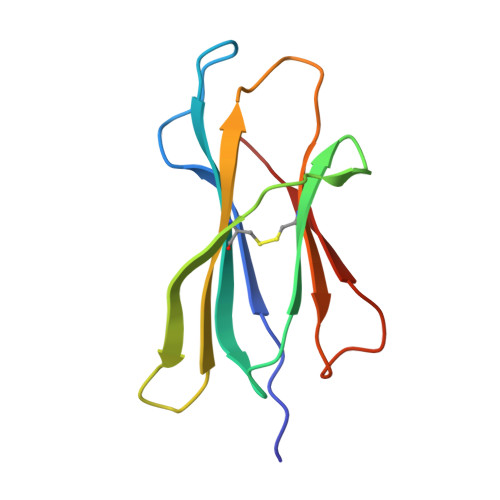
A structural basis for antigen presentation by the MHC class Ib molecule, Qa-1b

Zeng, L.Sullivan, L.C.Vivian, J.P.Walpole, N.G.Harpur, C.M.Rossjohn, J.Clements, C.S.Brooks, A.G.

(2012) J Immunol 188: 302-310

  • DOI: https://doi.org/10.4049/jimmunol.1102379
  • Primary Citation of Related Structures:  
    3VJ6

  • PubMed Abstract: 

    The primary function of the monomorphic MHC class Ib molecule Qa-1(b) is to present peptides derived from the leader sequences of other MHC class I molecules for recognition by the CD94-NKG2 receptors expressed by NK and T cells. Whereas the mode of peptide presentation by its ortholog HLA-E, and subsequent recognition by CD94-NKG2A, is known, the molecular basis of Qa-1(b) function is unclear. We have assessed the interaction between Qa-1(b) and CD94-NKG2A and shown that they interact with an affinity of 17 μM. Furthermore, we have determined the structure of Qa-1(b) bound to the leader sequence peptide, Qdm (AMAPRTLLL), to a resolution of 1.9 Å and compared it with that of HLA-E. The crystal structure provided a basis for understanding the restricted peptide repertoire of Qa-1(b). Whereas the Qa-1(b-AMAPRTLLL) complex was similar to that of HLA-E, significant sequence and structural differences were observed between the respective Ag-binding clefts. However, the conformation of the Qdm peptide bound by Qa-1(b) was very similar to that of peptide bound to HLA-E. Although a number of conserved innate receptors can recognize heterologous ligands from other species, the structural differences between Qa-1(b) and HLA-E manifested in CD94-NKG2A ligand recognition being species specific despite similarities in peptide sequence and conformation. Collectively, our data illustrate the structural homology between Qa-1(b) and HLA-E and provide a structural basis for understanding peptide repertoire selection and the specificity of the interaction of Qa-1(b) with CD94-NKG2 receptors.


  • Organizational Affiliation

    Department of Biochemistry and Molecular Biology, Monash University, Clayton, Victoria, Australia.


Macromolecules
Find similar proteins by:  (by identity cutoff)  |  3D Structure
Entity ID: 1
MoleculeChains Sequence LengthOrganismDetailsImage
H-2 class I histocompatibility antigen, D-37 alpha chain277Mus musculusMutation(s): 0 
Gene Names: H2-T23
UniProt
Find proteins for P06339 (Mus musculus)
Explore P06339 
Go to UniProtKB:  P06339
Entity Groups  
Sequence Clusters30% Identity50% Identity70% Identity90% Identity95% Identity100% Identity
UniProt GroupP06339
Sequence Annotations
Expand
  • Reference Sequence
Find similar proteins by:  (by identity cutoff)  |  3D Structure
Entity ID: 2
MoleculeChains Sequence LengthOrganismDetailsImage
Beta-2-microglobulin100Mus musculusMutation(s): 0 
Gene Names: B2m
UniProt & NIH Common Fund Data Resources
Find proteins for P01887 (Mus musculus)
Explore P01887 
Go to UniProtKB:  P01887
IMPC:  MGI:88127
Entity Groups  
Sequence Clusters30% Identity50% Identity70% Identity90% Identity95% Identity100% Identity
UniProt GroupP01887
Sequence Annotations
Expand
  • Reference Sequence

Find similar proteins by:  Sequence   |   3D Structure  

Entity ID: 3
MoleculeChains Sequence LengthOrganismDetailsImage
Qdm peptideC [auth P]9Mus musculusMutation(s): 0 
UniProt
Find proteins for P01897 (Mus musculus)
Explore P01897 
Go to UniProtKB:  P01897
Entity Groups  
UniProt GroupP01897
Sequence Annotations
Expand
  • Reference Sequence
Experimental Data & Validation

Experimental Data

  • Method: X-RAY DIFFRACTION
  • Resolution: 1.90 Å
  • R-Value Free: 0.242 
  • R-Value Work: 0.201 
  • R-Value Observed: 0.203 
  • Space Group: P 21 21 21
Unit Cell:
Length ( Å )Angle ( ˚ )
a = 59.268α = 90
b = 72.833β = 90
c = 95.844γ = 90
Software Package:
Software NamePurpose
CrystalCleardata collection
PHASERphasing
PHENIXrefinement
HKL-2000data reduction
SCALEPACKdata scaling

Structure Validation

View Full Validation Report


Entry History 

Deposition Data

Revision History  (Full details and data files)

  • Version 1.0: 2012-03-14
    Type: Initial release
  • Version 1.1: 2023-11-08
    Changes: Data collection, Database references, Derived calculations, Refinement description