3UTN

Crystal structure of Tum1 protein from Saccharomyces cerevisiae


Experimental Data Snapshot

  • Method: X-RAY DIFFRACTION
  • Resolution: 1.90 Å
  • R-Value Free: 0.228 
  • R-Value Work: 0.204 
  • R-Value Observed: 0.205 

wwPDB Validation   3D Report Full Report


This is version 1.4 of the entry. See complete history


Literature

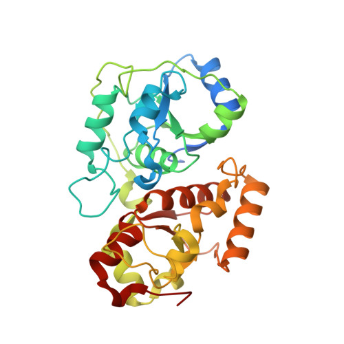
Crystal structure of the Tum1 protein from the yeast Saccharomyces cerevisiae.

Qiu, R.Wang, F.Liu, M.Lou, T.Ji, C.

(2012) Protein Pept Lett 19: 1139-1143

  • DOI: https://doi.org/10.2174/092986612803217060
  • Primary Citation of Related Structures:  
    3UTN

  • PubMed Abstract: 

    Yeast tRNA-thiouridine modification protein 1 (Tum1) plays essential role in the sulfur transfer process of Urm1 system, which in turn is involved in many important cellular processes. In the rhodanese-like domain (RLD), conserved cysteine residue is proved to be the centre of active site of sulfurtransferases and crucial for the substrate recognition. In this report, we describe the crystal structure of Tum1 protein at 1.90 A resolution which, despite consisting of two RLDs, has only one conserved cysteine residue in the C-terminal RLD. An unaccounted electron density is found near the active site, which might point to the new cofactor in the sulfur transfer mechanism.


  • Organizational Affiliation

    State Key Laboratory of Genetic Engineering, Institute of Genetics, School of Life Sciences, Fudan University, Shanghai, People’s Republic of China.


Macromolecules
Find similar proteins by:  (by identity cutoff)  |  3D Structure
Entity ID: 1
MoleculeChains Sequence LengthOrganismDetailsImage
Thiosulfate sulfurtransferase TUM1A [auth X]327Saccharomyces cerevisiae S288CMutation(s): 0 
Gene Names: TUM1YOR251C
EC: 2.8.1.1
UniProt
Find proteins for Q08686 (Saccharomyces cerevisiae (strain ATCC 204508 / S288c))
Explore Q08686 
Go to UniProtKB:  Q08686
Entity Groups  
Sequence Clusters30% Identity50% Identity70% Identity90% Identity95% Identity100% Identity
UniProt GroupQ08686
Sequence Annotations
Expand
  • Reference Sequence
Small Molecules
Ligands 2 Unique
IDChains Name / Formula / InChI Key2D Diagram3D Interactions
SO4
Query on SO4

Download Ideal Coordinates CCD File 
K [auth X]SULFATE ION
O4 S
QAOWNCQODCNURD-UHFFFAOYSA-L
DMS
Query on DMS

Download Ideal Coordinates CCD File 
B [auth X]
C [auth X]
D [auth X]
E [auth X]
F [auth X]
B [auth X],
C [auth X],
D [auth X],
E [auth X],
F [auth X],
G [auth X],
H [auth X],
I [auth X],
J [auth X]
DIMETHYL SULFOXIDE
C2 H6 O S
IAZDPXIOMUYVGZ-UHFFFAOYSA-N
Experimental Data & Validation

Experimental Data

  • Method: X-RAY DIFFRACTION
  • Resolution: 1.90 Å
  • R-Value Free: 0.228 
  • R-Value Work: 0.204 
  • R-Value Observed: 0.205 
  • Space Group: I 41
Unit Cell:
Length ( Å )Angle ( ˚ )
a = 120.939α = 90
b = 120.939β = 90
c = 48.354γ = 90
Software Package:
Software NamePurpose
DENZOdata reduction
SCALEPACKdata scaling
PHASERphasing
PHENIXrefinement
PDB_EXTRACTdata extraction
HKL-2000data collection
HKL-2000data reduction
HKL-2000data scaling
PHASESphasing
REFMACrefinement

Structure Validation

View Full Validation Report


Entry History 

Deposition Data

Revision History  (Full details and data files)

  • Version 1.0: 2012-10-17
    Type: Initial release
  • Version 1.1: 2013-07-17
    Changes: Database references
  • Version 1.2: 2017-11-08
    Changes: Refinement description
  • Version 1.3: 2019-10-09
    Changes: Data collection, Database references, Derived calculations
  • Version 1.4: 2023-11-08
    Changes: Data collection, Database references, Derived calculations, Refinement description