Protonation-state determination in proteins using high-resolution X-ray crystallography: effects of resolution and completeness.
Fisher, S.J., Blakeley, M.P., Cianci, M., McSweeney, S., Helliwell, J.R.(2012) Acta Crystallogr D Biol Crystallogr 68: 800-809
- PubMed: 22751665
- DOI: https://doi.org/10.1107/S0907444912012589
- Primary Citation of Related Structures:
3UNX, 4YTA - PubMed Abstract:
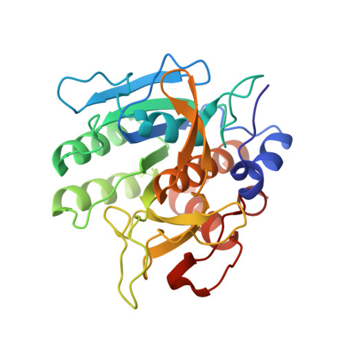
A bond-distance analysis has been undertaken to determine the protonation states of ionizable amino acids in trypsin, subtilisin and lysozyme. The diffraction resolutions were 1.2 Å for trypsin (97% complete, 12% H-atom visibility at 2.5σ), 1.26 Å for subtilisin (100% complete, 11% H-atom visibility at 2.5σ) and 0.65 Å for lysozyme (PDB entry 2vb1; 98% complete, 30% H-atom visibility at 3σ). These studies provide a wide diffraction resolution range for assessment. The bond-length e.s.d.s obtained are as small as 0.008 Å and thus provide an exceptional opportunity for bond-length analyses. The results indicate that useful information can be obtained from diffraction data at around 1.2-1.3 Å resolution and that minor increases in resolution can have significant effects on reducing the associated bond-length standard deviations. The protonation states in histidine residues were also considered; however, owing to the smaller differences between the protonated and deprotonated forms it is much more difficult to infer the protonation states of these residues. Not even the 0.65 Å resolution lysozyme structure provided the necessary accuracy to determine the protonation states of histidine.
Organizational Affiliation:
School of Chemistry, University of Manchester, Brunswick Street, Manchester M13 9PL, England. fisher@ill.fr


