3ULK

E. coli Ketol-acid reductoisomerase in complex with NADPH and Mg2+


Experimental Data Snapshot

  • Method: X-RAY DIFFRACTION
  • Resolution: 2.30 Å
  • R-Value Free: 0.236 
  • R-Value Work: 0.174 
  • R-Value Observed: 0.177 

wwPDB Validation   3D Report Full Report


Ligand Structure Quality Assessment 


This is version 1.2 of the entry. See complete history


Literature

Bacterial and plant ketol-acid reductoisomerases have different mechanisms of induced fit during the catalytic cycle

Wong, S.H.Lonhienne, T.G.A.Winzor, D.J.Schenk, G.Guddat, L.W.

(2012) J Mol Biol 424: 168-179

  • DOI: https://doi.org/10.1016/j.jmb.2012.09.018
  • Primary Citation of Related Structures:  
    3ULK

  • PubMed Abstract: 

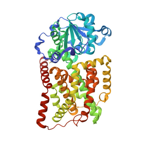
    Ketol-acid reductoisomerase (KARI) is the second enzyme in the branched-chain amino acid biosynthesis pathway, which is found in plants, fungi and bacteria but not in animals. This difference in metabolism between animals and microorganisms makes KARI an attractive target for the development of antimicrobial agents. Herein we report the crystal structure of Escherichia coli KARI in complex with Mg(2+) and NADPH at 2.3Å resolution. Ultracentrifugation studies confirm that the enzyme exists as a tetramer in solution, and isothermal titration calorimetry shows that the binding of Mg(2+) increases structural disorder while the binding of NADPH increases the structural rigidity of the enzyme. Comparison of the structure of the E. coli KARI-Mg(2+)-NADPH complex with that of enzyme in the absence of cofactors shows that the binding of Mg(2+) and NADPH opens the interface between the N- and C-domains, thereby allowing access for the substrates to bind: the existence of only a small opening between the domains in the crystal structure of the unliganded enzyme signifies restricted access to the active site. This observation contrasts with that in the plant enzyme, where the N-domain can rotate freely with respect to the C-domain until the binding of Mg(2+) and/or NADPH stabilizes the relative positions of these domains. Support is thereby provided for the idea that plant and bacterial KARIs have evolved different mechanisms of induced fit to prepare the active site for catalysis.


  • Organizational Affiliation

    School of Chemistry and Molecular Biosciences, The University of Queensland, Brisbane, Queensland 4072, Australia. s.wong3@uq.edu.au


Macromolecules
Find similar proteins by:  (by identity cutoff)  |  3D Structure
Entity ID: 1
MoleculeChains Sequence LengthOrganismDetailsImage
Ketol-acid reductoisomerase
A, B
491Escherichia coli K-12Mutation(s): 0 
Gene Names: ilvC
EC: 1.1.1.86
UniProt
Find proteins for P05793 (Escherichia coli (strain K12))
Explore P05793 
Go to UniProtKB:  P05793
Entity Groups  
Sequence Clusters30% Identity50% Identity70% Identity90% Identity95% Identity100% Identity
UniProt GroupP05793
Sequence Annotations
Expand
  • Reference Sequence
Small Molecules
Ligands 3 Unique
IDChains Name / Formula / InChI Key2D Diagram3D Interactions
NDP
Query on NDP

Download Ideal Coordinates CCD File 
C [auth A],
K [auth B]
NADPH DIHYDRO-NICOTINAMIDE-ADENINE-DINUCLEOTIDE PHOSPHATE
C21 H30 N7 O17 P3
ACFIXJIJDZMPPO-NNYOXOHSSA-N
SO4
Query on SO4

Download Ideal Coordinates CCD File 
D [auth A]
E [auth A]
F [auth A]
G [auth A]
H [auth A]
D [auth A],
E [auth A],
F [auth A],
G [auth A],
H [auth A],
L [auth B],
M [auth B],
N [auth B],
O [auth B]
SULFATE ION
O4 S
QAOWNCQODCNURD-UHFFFAOYSA-L
MG
Query on MG

Download Ideal Coordinates CCD File 
I [auth A],
J [auth A],
P [auth B],
Q [auth B]
MAGNESIUM ION
Mg
JLVVSXFLKOJNIY-UHFFFAOYSA-N
Modified Residues  1 Unique
IDChains TypeFormula2D DiagramParent
CSX
Query on CSX
A, B
L-PEPTIDE LINKINGC3 H7 N O3 SCYS
Binding Affinity Annotations 
IDSourceBinding Affinity
NDP Binding MOAD:  3ULK Kd: 1.93e+4 (nM) from 1 assay(s)
Experimental Data & Validation

Experimental Data

  • Method: X-RAY DIFFRACTION
  • Resolution: 2.30 Å
  • R-Value Free: 0.236 
  • R-Value Work: 0.174 
  • R-Value Observed: 0.177 
  • Space Group: P 64 2 2
Unit Cell:
Length ( Å )Angle ( ˚ )
a = 144.459α = 90
b = 144.459β = 90
c = 217.545γ = 120
Software Package:
Software NamePurpose
ADSCdata collection
MOLREPphasing
PHENIXrefinement
HKL-2000data reduction
HKL-2000data scaling

Structure Validation

View Full Validation Report


Ligand Structure Quality Assessment 


Entry History 

Deposition Data

Revision History  (Full details and data files)

  • Version 1.0: 2012-10-17
    Type: Initial release
  • Version 1.1: 2013-06-26
    Changes: Database references
  • Version 1.2: 2023-11-01
    Changes: Data collection, Database references, Derived calculations, Refinement description