Structural and functional characterization of solute binding proteins for aromatic compounds derived from lignin: p-Coumaric acid and related aromatic acids.
Tan, K., Chang, C., Cuff, M., Osipiuk, J., Landorf, E., Mack, J.C., Zerbs, S., Joachimiak, A., Collart, F.R.(2013) Proteins 81: 1709-1726
- PubMed: 23606130
- DOI: https://doi.org/10.1002/prot.24305
- Primary Citation of Related Structures:
3RPW, 3SG0, 3TX6, 3UK0, 3UKJ, 4DQD, 4EYO, 4EYQ, 4F8J, 4FB4, 4I1D - PubMed Abstract:
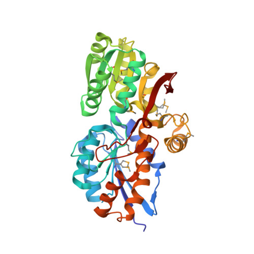
Lignin comprises 15-25% of plant biomass and represents a major environmental carbon source for utilization by soil microorganisms. Access to this energy resource requires the action of fungal and bacterial enzymes to break down the lignin polymer into a complex assortment of aromatic compounds that can be transported into the cells. To improve our understanding of the utilization of lignin by microorganisms, we characterized the molecular properties of solute binding proteins of ATP-binding cassette transporter proteins that interact with these compounds. A combination of functional screens and structural studies characterized the binding specificity of the solute binding proteins for aromatic compounds derived from lignin such as p-coumarate, 3-phenylpropionic acid and compounds with more complex ring substitutions. A ligand screen based on thermal stabilization identified several binding protein clusters that exhibit preferences based on the size or number of aromatic ring substituents. Multiple X-ray crystal structures of protein-ligand complexes for these clusters identified the molecular basis of the binding specificity for the lignin-derived aromatic compounds. The screens and structural data provide new functional assignments for these solute-binding proteins which can be used to infer their transport specificity. This knowledge of the functional roles and molecular binding specificity of these proteins will support the identification of the specific enzymes and regulatory proteins of peripheral pathways that funnel these compounds to central metabolic pathways and will improve the predictive power of sequence-based functional annotation methods for this family of proteins.
Organizational Affiliation:
Biosciences Division, Argonne National Laboratory, Lemont, Illinois, 60439; The Midwest Center for Structural Genomics, Argonne National Laboratory, Lemont, Illinois, 60439; Structural Biology Center, Argonne National Laboratory, Lemont, Illinois, 60439.


