Structural study of TTR-52 reveals the mechanism by which a bridging molecule mediates apoptotic cell engulfment
Kang, Y.Y., Zhao, D.F., Liang, H.H., Liu, B., Zhang, Y., Liu, Q.W., Wang, X.C., Liu, Y.F.(2012) Genes Dev 26: 1339-1350
- PubMed: 22713871
- DOI: https://doi.org/10.1101/gad.187815.112
- Primary Citation of Related Structures:
3UAF - PubMed Abstract:
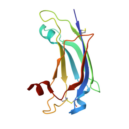
During apoptosis, apoptotic cells are removed by professional phagocytes or neighboring engulfing cells either directly through phagocytic receptors or indirectly through bridging molecules that cross-link dying cells to phagocytes. However, how bridging molecules recognize "eat me" signals and phagocytic receptors to mediate engulfment remains unclear. Here, we report the structural and functional studies of Caenorhabditis elegans TTR-52, a recently identified bridging molecule that cross-links surface-exposed phosphatidylserine (PtdSer) on apoptotic cells to the CED-1 receptor on phagocytes. Crystal structure studies show that TTR-52 has an open β-barrel-like structure with some similarities to the PKCα-C2 domain. TTR-52 is proposed to bind PtdSer via an "ion-mediating" PtdSer-binding mode. Intensive functional studies show that CED-1 binds TTR-52 through its N-terminal EMI domain and that the hydrophobic region of the TTR-52 C terminus is involved in this interaction. In addition, unlike other PtdSer-binding domains, TTR-52 forms dimers, and its dimerization is important for its function in vivo. Our results reveal the first full-length structure of a bridging molecule and the mechanism underlying bridging molecule-mediated apoptotic cell recognition.
Organizational Affiliation:
State Key Laboratory of Biomacromolecules, Institute of Biophysics, Chinese Academy of Sciences, Chaoyang District, Beijing 100101, China.