3U9P

Crystal Structure of Murine Siderocalin in Complex with an Fab Fragment


Experimental Data Snapshot

  • Method: X-RAY DIFFRACTION
  • Resolution: 2.80 Å
  • R-Value Free: 0.292 
  • R-Value Work: 0.219 
  • R-Value Observed: 0.223 

wwPDB Validation   3D Report Full Report


This is version 1.2 of the entry. See complete history


Literature

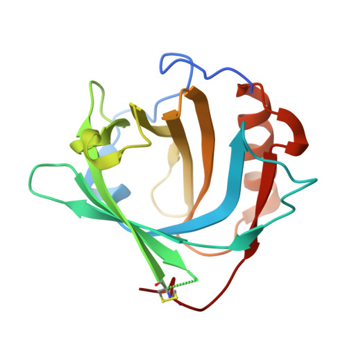
Siderocalin/Lcn2/NGAL/24p3 does not drive apoptosis through gentisic acid mediated iron withdrawal in hematopoietic cell lines.

Correnti, C.Richardson, V.Sia, A.K.Bandaranayake, A.D.Ruiz, M.Suryo Rahmanto, Y.Kovacevic, Z.Clifton, M.C.Holmes, M.A.Kaiser, B.K.Barasch, J.Raymond, K.N.Richardson, D.R.Strong, R.K.

(2012) PLoS One 7: e43696-e43696

  • DOI: https://doi.org/10.1371/journal.pone.0043696
  • Primary Citation of Related Structures:  
    3U0D, 3U9P

  • PubMed Abstract: 

    Siderocalin (also lipocalin 2, NGAL or 24p3) binds iron as complexes with specific siderophores, which are low molecular weight, ferric ion-specific chelators. In innate immunity, siderocalin slows the growth of infecting bacteria by sequestering bacterial ferric siderophores. Siderocalin also binds simple catechols, which can serve as siderophores in the damaged urinary tract. Siderocalin has also been proposed to alter cellular iron trafficking, for instance, driving apoptosis through iron efflux via BOCT. An endogenous siderophore composed of gentisic acid (2,5-dihydroxybenzoic acid) substituents was proposed to mediate cellular efflux. However, binding studies reported herein contradict the proposal that gentisic acid forms high-affinity ternary complexes with siderocalin and iron, or that gentisic acid can serve as an endogenous siderophore at neutral pH. We also demonstrate that siderocalin does not induce cellular iron efflux or stimulate apoptosis, questioning the role siderocalin plays in modulating iron metabolism.


  • Organizational Affiliation

    Division of Basic Sciences, Fred Hutchinson Cancer Research Center, Seattle, Washington, United States of America.


Macromolecules
Find similar proteins by:  (by identity cutoff)  |  3D Structure
Entity ID: 1
MoleculeChains Sequence LengthOrganismDetailsImage
Neutrophil gelatinase-associated lipocalinA [auth C],
B [auth D]
182Mus musculusMutation(s): 0 
Gene Names: Lcn2
UniProt & NIH Common Fund Data Resources
Find proteins for P11672 (Mus musculus)
Explore P11672 
Go to UniProtKB:  P11672
IMPC:  MGI:96757
Entity Groups  
Sequence Clusters30% Identity50% Identity70% Identity90% Identity95% Identity100% Identity
UniProt GroupP11672
Sequence Annotations
Expand
  • Reference Sequence
Find similar proteins by:  (by identity cutoff)  |  3D Structure
Entity ID: 2
MoleculeChains Sequence LengthOrganismDetailsImage
Monoclonal Fab Fragment Heavy ChainC [auth H],
D [auth K]
216Rattus norvegicusMutation(s): 0 
Entity Groups  
Sequence Clusters30% Identity50% Identity70% Identity90% Identity95% Identity100% Identity
Sequence Annotations
Expand
  • Reference Sequence
Find similar proteins by:  (by identity cutoff)  |  3D Structure
Entity ID: 3
MoleculeChains Sequence LengthOrganismDetailsImage
Monoclonal Fab Fragment Light ChainE [auth L],
F [auth M]
212Rattus norvegicusMutation(s): 0 
Entity Groups  
Sequence Clusters30% Identity50% Identity70% Identity90% Identity95% Identity100% Identity
Sequence Annotations
Expand
  • Reference Sequence
Experimental Data & Validation

Experimental Data

  • Method: X-RAY DIFFRACTION
  • Resolution: 2.80 Å
  • R-Value Free: 0.292 
  • R-Value Work: 0.219 
  • R-Value Observed: 0.223 
  • Space Group: P 21 21 21
Unit Cell:
Length ( Å )Angle ( ˚ )
a = 82.133α = 90
b = 124.091β = 90
c = 147.487γ = 90
Software Package:
Software NamePurpose
DENZOdata reduction
SCALEPACKdata scaling
REFMACrefinement
PDB_EXTRACTdata extraction
ADSCdata collection
HKL-2000data reduction
PHASERphasing

Structure Validation

View Full Validation Report


Entry History 

Deposition Data

Revision History  (Full details and data files)

  • Version 1.0: 2013-05-01
    Type: Initial release
  • Version 1.1: 2017-11-08
    Changes: Refinement description
  • Version 1.2: 2018-01-31
    Changes: Database references