3TUR

Crystal Structure of M. tuberculosis LD-transpeptidase type 2 complexed with a peptidoglycan fragment


Experimental Data Snapshot

  • Method: X-RAY DIFFRACTION
  • Resolution: 1.72 Å
  • R-Value Free: 0.235 
  • R-Value Work: 0.199 
  • R-Value Observed: 0.200 

wwPDB Validation   3D Report Full Report


Ligand Structure Quality Assessment 


This is version 1.4 of the entry. See complete history


Literature

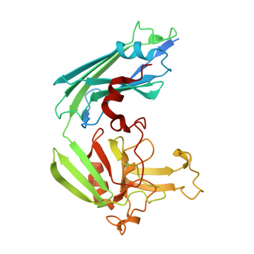
Targeting the Cell Wall of Mycobacterium tuberculosis: Structure and Mechanism of L,D-Transpeptidase 2.

Erdemli, S.B.Gupta, R.Bishai, W.R.Lamichhane, G.Amzel, L.M.Bianchet, M.A.

(2012) Structure 20: 2103-2115

  • DOI: https://doi.org/10.1016/j.str.2012.09.016
  • Primary Citation of Related Structures:  
    3TUR, 3TX4, 3U1P, 3VAE

  • PubMed Abstract: 

    With multidrug-resistant cases of tuberculosis increasing globally, better antibiotic drugs and novel drug targets are becoming an urgent need. Traditional β-lactam antibiotics that inhibit D,D-transpeptidases are not effective against mycobacteria, in part because mycobacteria rely mostly on L,D-transpeptidases for biosynthesis and maintenance of their peptidoglycan layer. This reliance plays a major role in drug resistance and persistence of Mycobacterium tuberculosis (Mtb) infections. The crystal structure at 1.7 Å resolution of the Mtb L,D-transpeptidase Ldt(Mt2) containing a bound peptidoglycan fragment, reported here, provides information about catalytic site organization as well as substrate recognition by the enzyme. Based on our structural, kinetic, and calorimetric data, we propose a catalytic mechanism for Ldt(Mt2) in which both acyl-acceptor and acyl-donor substrates reach the catalytic site from the same, rather than different, entrances. Together, this information provides vital insights to facilitate development of drugs targeting this validated yet unexploited enzyme.


  • Organizational Affiliation

    Department of Biophysics and Biophysical Chemistry, Johns Hopkins University School of Medicine, Baltimore, MD 21205, USA.


Macromolecules
Find similar proteins by:  (by identity cutoff)  |  3D Structure
Entity ID: 1
MoleculeChains Sequence LengthOrganismDetailsImage
Mycobacteria Tuberculosis LD-transpeptidase type 2
A, B
287Mycobacterium tuberculosisMutation(s): 0 
Gene Names: lppSMT2594Rv2518c
UniProt
Find proteins for O53223 (Mycobacterium tuberculosis (strain CDC 1551 / Oshkosh))
Explore O53223 
Go to UniProtKB:  O53223
Entity Groups  
Sequence Clusters30% Identity50% Identity70% Identity90% Identity95% Identity100% Identity
UniProt GroupO53223
Sequence Annotations
Expand
  • Reference Sequence
Small Molecules
Ligands 4 Unique
IDChains Name / Formula / InChI Key2D Diagram3D Interactions
0JC
Query on 0JC

Download Ideal Coordinates CCD File 
C [auth A]
D [auth A]
E [auth A]
F [auth A]
J [auth B]
C [auth A],
D [auth A],
E [auth A],
F [auth A],
J [auth B],
K [auth B],
L [auth B],
M [auth B]
Di-mu-iodobis(ethylenediamine)diplatinum(II)
C4 H16 I2 N4 Pt2
XXLKNYJQAYMQHB-UHFFFAOYSA-N
PT
Query on PT

Download Ideal Coordinates CCD File 
I [auth B]PLATINUM (II) ION
Pt
HRGDZIGMBDGFTC-UHFFFAOYSA-N
6CL
Query on 6CL

Download Ideal Coordinates CCD File 
H [auth A]6-CARBOXYLYSINE
C7 H15 N2 O4
GMKMEZVLHJARHF-SYDPRGILSA-O
DGL
Query on DGL

Download Ideal Coordinates CCD File 
G [auth A]D-GLUTAMIC ACID
C5 H9 N O4
WHUUTDBJXJRKMK-GSVOUGTGSA-N
Experimental Data & Validation

Experimental Data

  • Method: X-RAY DIFFRACTION
  • Resolution: 1.72 Å
  • R-Value Free: 0.235 
  • R-Value Work: 0.199 
  • R-Value Observed: 0.200 
  • Space Group: I 21 21 21
Unit Cell:
Length ( Å )Angle ( ˚ )
a = 119.132α = 90
b = 120.829β = 90
c = 122.847γ = 90
Software Package:
Software NamePurpose
REFMACrefinement
PDB_EXTRACTdata extraction
HKL-2000data collection
DENZOdata reduction
SCALEPACKdata scaling
SHARPphasing

Structure Validation

View Full Validation Report


Ligand Structure Quality Assessment 


Entry History 

Deposition Data

Revision History  (Full details and data files)

  • Version 1.0: 2012-12-05
    Type: Initial release
  • Version 1.1: 2012-12-12
    Changes: Database references
  • Version 1.2: 2012-12-26
    Changes: Database references
  • Version 1.3: 2017-11-08
    Changes: Advisory, Refinement description
  • Version 1.4: 2024-02-28
    Changes: Data collection, Database references, Derived calculations