


Crystal structure of putative aminomethyltransferase from Leptospirillum rubarum
Michalska, K., Xu, X., Cui, H., Chin, S., Savchenko, A., Joachimiak, A.To be published.
Experimental Data Snapshot
wwPDB Validation 3D Report Full Report

Entity ID: 1 | |||||
|---|---|---|---|---|---|
| Molecule | Chains | Sequence Length | Organism | Details | Image |
| Putative aminomethyltransferase | 355 | Leptospirillum rubarum | Mutation(s): 0 Gene Names: EAY58150, UBAL2_82410567 | 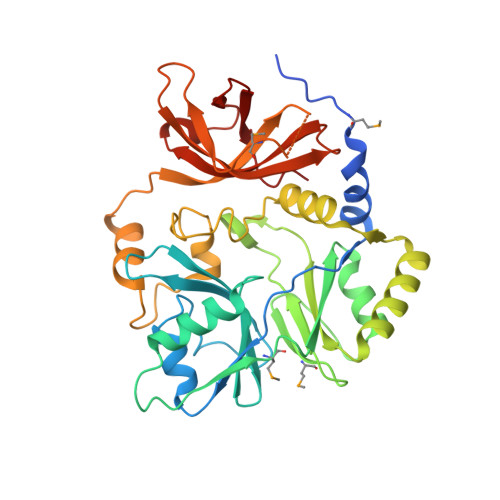 | |
Entity Groups | |||||
| Sequence Clusters | 30% Identity50% Identity70% Identity90% Identity95% Identity100% Identity | ||||
Sequence AnnotationsExpand | |||||
| |||||
| Ligands 1 Unique | |||||
|---|---|---|---|---|---|
| ID | Chains | Name / Formula / InChI Key | 2D Diagram | 3D Interactions | |
| CL Query on CL | B [auth A], C [auth A], D [auth A], E [auth A] | CHLORIDE ION Cl VEXZGXHMUGYJMC-UHFFFAOYSA-M |  | ||
| Modified Residues 1 Unique | |||||
|---|---|---|---|---|---|
| ID | Chains | Type | Formula | 2D Diagram | Parent |
| MSE Query on MSE | A | L-PEPTIDE LINKING | C5 H11 N O2 Se |  | MET |
| Length ( Å ) | Angle ( ˚ ) |
|---|---|
| a = 115.616 | α = 90 |
| b = 115.616 | β = 90 |
| c = 75.428 | γ = 90 |
| Software Name | Purpose |
|---|---|
| SBC-Collect | data collection |
| SHELX | model building |
| MLPHARE | phasing |
| DM | model building |
| ARP/wARP | model building |
| Coot | model building |
| BUSTER | refinement |
| HKL-3000 | data reduction |
| HKL-3000 | data scaling |
| SHELX | phasing |
| DM | phasing |
RCSB PDB (citation) is hosted by
RCSB PDB is a member of the

