3TMA

Crystal structure of TrmN from Thermus thermophilus


Experimental Data Snapshot

  • Method: X-RAY DIFFRACTION
  • Resolution: 2.05 Å
  • R-Value Free: 0.212 
  • R-Value Work: 0.154 
  • R-Value Observed: 0.157 

wwPDB Validation   3D Report Full Report


This is version 1.3 of the entry. See complete history


Literature

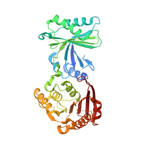
Crystal structures of the tRNA:m2G6 methyltransferase Trm14/TrmN from two domains of life.

Fislage, M.Roovers, M.Tuszynska, I.Bujnicki, J.M.Droogmans, L.Versees, W.

(2012) Nucleic Acids Res 40: 5149-5161

  • DOI: https://doi.org/10.1093/nar/gks163
  • Primary Citation of Related Structures:  
    3TLJ, 3TM4, 3TM5, 3TMA

  • PubMed Abstract: 

    Methyltransferases (MTases) form a major class of tRNA-modifying enzymes needed for the proper functioning of tRNA. Recently, RNA MTases from the TrmN/Trm14 family that are present in Archaea, Bacteria and Eukaryota have been shown to specifically modify tRNA(Phe) at guanosine 6 in the tRNA acceptor stem. Here, we report the first X-ray crystal structures of the tRNA m(2)G6 (N(2)-methylguanosine) MTase (TTC)TrmN from Thermus thermophilus and its ortholog (Pf)Trm14 from Pyrococcus furiosus. Structures of (Pf)Trm14 were solved in complex with the methyl donor S-adenosyl-l-methionine (SAM or AdoMet), as well as the reaction product S-adenosyl-homocysteine (SAH or AdoHcy) and the inhibitor sinefungin. (TTC)TrmN and (Pf)Trm14 consist of an N-terminal THUMP domain fused to a catalytic Rossmann-fold MTase (RFM) domain. These results represent the first crystallographic structure analysis of proteins containing both THUMP and RFM domain, and hence provide further insight in the contribution of the THUMP domain in tRNA recognition and catalysis. Electrostatics and conservation calculations suggest a main tRNA binding surface in a groove between the THUMP domain and the MTase domain. This is further supported by a docking model of TrmN in complex with tRNA(Phe) of T. thermophilus and via site-directed mutagenesis.


  • Organizational Affiliation

    VIB Department of Structural Biology, Vrije Universiteit Brussel, Pleinlaan 2, 1050 Brussel, Belgium.


Macromolecules
Find similar proteins by:  (by identity cutoff)  |  3D Structure
Entity ID: 1
MoleculeChains Sequence LengthOrganismDetailsImage
methyltransferase354Thermus thermophilus HB27Mutation(s): 0 
Gene Names: TTC1157TT_C1157
EC: 2.1.1
UniProt
Find proteins for Q72IH5 (Thermus thermophilus (strain ATCC BAA-163 / DSM 7039 / HB27))
Explore Q72IH5 
Go to UniProtKB:  Q72IH5
Entity Groups  
Sequence Clusters30% Identity50% Identity70% Identity90% Identity95% Identity100% Identity
UniProt GroupQ72IH5
Sequence Annotations
Expand
  • Reference Sequence
Small Molecules
Ligands 1 Unique
IDChains Name / Formula / InChI Key2D Diagram3D Interactions
PO4
Query on PO4

Download Ideal Coordinates CCD File 
B [auth A]PHOSPHATE ION
O4 P
NBIIXXVUZAFLBC-UHFFFAOYSA-K
Experimental Data & Validation

Experimental Data

  • Method: X-RAY DIFFRACTION
  • Resolution: 2.05 Å
  • R-Value Free: 0.212 
  • R-Value Work: 0.154 
  • R-Value Observed: 0.157 
  • Space Group: P 32 2 1
Unit Cell:
Length ( Å )Angle ( ˚ )
a = 65.86α = 90
b = 65.86β = 90
c = 144.55γ = 120
Software Package:
Software NamePurpose
REFMACrefinement

Structure Validation

View Full Validation Report


Entry History 

Deposition Data

Revision History  (Full details and data files)

  • Version 1.0: 2012-03-14
    Type: Initial release
  • Version 1.1: 2012-05-09
    Changes: Other
  • Version 1.2: 2012-10-03
    Changes: Database references
  • Version 1.3: 2023-09-13
    Changes: Data collection, Database references, Derived calculations, Refinement description