3TKA

crystal structure and solution saxs of methyltransferase rsmh from E.coli


Experimental Data Snapshot

  • Method: X-RAY DIFFRACTION
  • Resolution: 2.25 Å
  • R-Value Free: 0.234 
  • R-Value Work: 0.184 
  • R-Value Observed: 0.187 

wwPDB Validation   3D Report Full Report


Ligand Structure Quality Assessment 


This is version 1.3 of the entry. See complete history


Literature

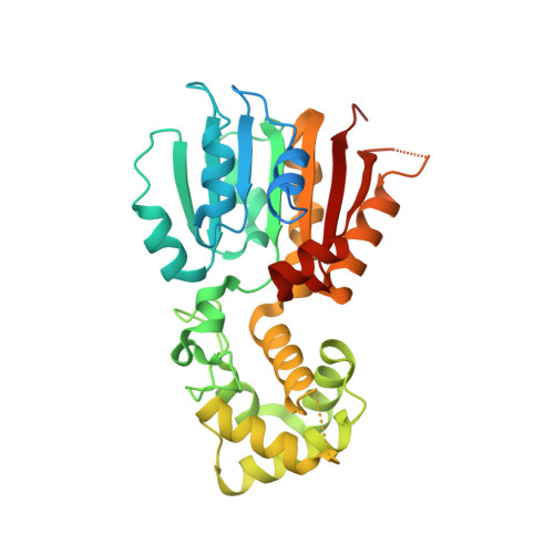
Crystal and solution structures of methyltransferase RsmH provide basis for methylation of C1402 in 16S rRNA.

Wei, Y.Zhang, H.Gao, Z.Q.Wang, W.J.Shtykova, E.V.Xu, J.H.Liu, Q.S.Dong, Y.H.

(2012) J Struct Biol 179: 29-40

  • DOI: https://doi.org/10.1016/j.jsb.2012.04.011
  • Primary Citation of Related Structures:  
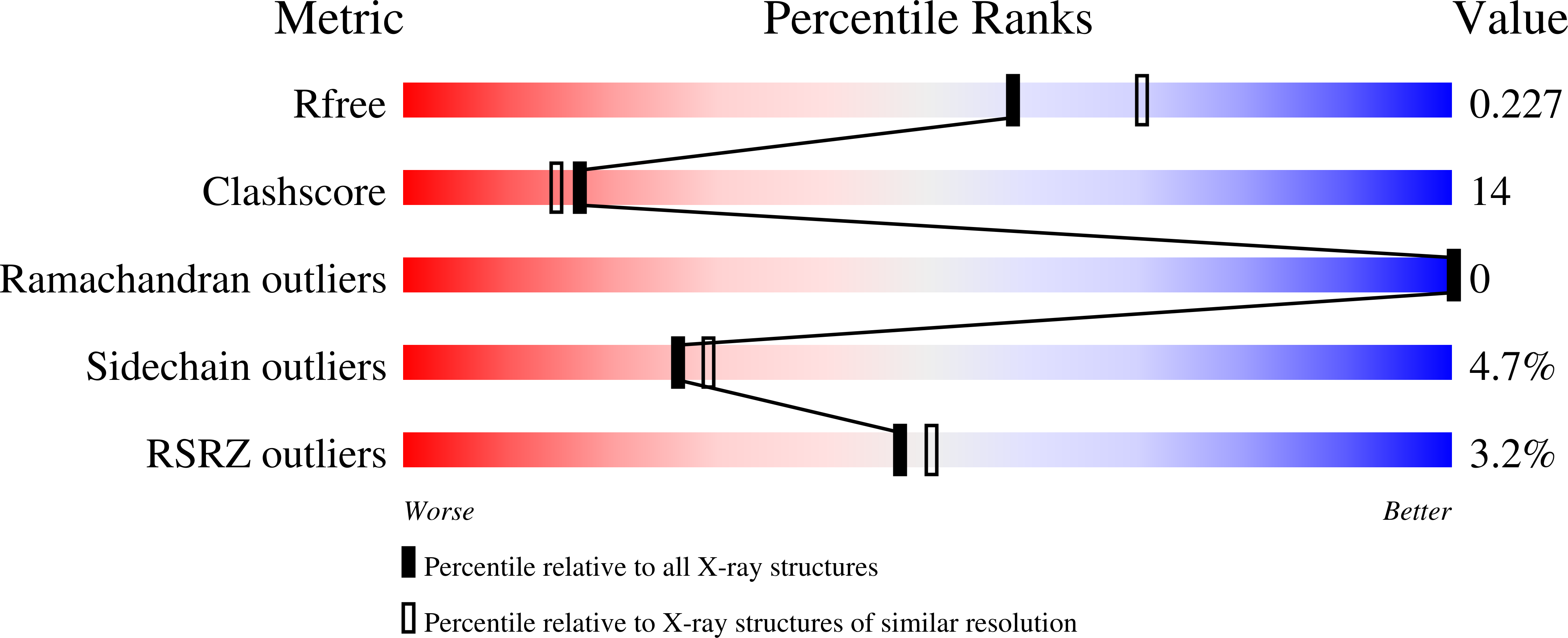
    3TKA

  • PubMed Abstract: 

    RsmH is a specific AdoMet-dependent methyltransferase (MTase) responsible for N(4)-methylation of C1402 in 16S rRNA and conserved in almost all species of bacteria. The methylcytidine interacts with the P-site codon of the mRNA and increases ribosomal decoding fidelity. In this study, high resolution crystal structure (2.25Å) of Escherichia coli RsmH in complex with AdoMet and cytidine (the putative rRNA binding site) was determined. The structural analysis demonstrated that the complex consists of two distinct but structurally related domains: the typical MTase domain and the putative substrate recognition and binding domain. A deep pocket was found in the conserved AdoMet binding domain. It was also found that the cytidine bound far from AdoMet with the distance of 25.9Å. It indicates that the complex is not in a catalytically active state, and structural rearrangement of RsmH or the nucleotides neighboring C1402 may be necessary to trigger catalysis. Although there is only one molecule in the asymmetric unit of the crystals, RsmH can form a compact dimer across a crystallographic twofold axis. Further analysis of RsmH by small-angle X-ray scattering (SAXS) also revealed the dimer in solution, but with a more flexible conformation than that in crystal, likely resulting from the absence of the substrate. It implies that an active status of RsmH in vivo is achieved by a formation of the dimeric architecture. In general, crystal and solution structural analysis provides new information on the mechanism of the methylation of the fine-tuning ribosomal decoding center by the RsmH.


  • Organizational Affiliation

    School of Life Sciences, University of Science and Technology of China, Hefei 230027, People's Republic of China.


Macromolecules
Find similar proteins by:  (by identity cutoff)  |  3D Structure
Entity ID: 1
MoleculeChains Sequence LengthOrganismDetailsImage
Ribosomal RNA small subunit methyltransferase H347Escherichia coli K-12Mutation(s): 0 
Gene Names: rsmHmraWyabCb0082JW0080
EC: 2.1.1.199
UniProt
Find proteins for P60390 (Escherichia coli (strain K12))
Explore P60390 
Go to UniProtKB:  P60390
Entity Groups  
Sequence Clusters30% Identity50% Identity70% Identity90% Identity95% Identity100% Identity
UniProt GroupP60390
Sequence Annotations
Expand
  • Reference Sequence
Small Molecules
Ligands 4 Unique
IDChains Name / Formula / InChI Key2D Diagram3D Interactions
SAM
Query on SAM

Download Ideal Coordinates CCD File 
B [auth A]S-ADENOSYLMETHIONINE
C15 H22 N6 O5 S
MEFKEPWMEQBLKI-FCKMPRQPSA-N
CTN
Query on CTN

Download Ideal Coordinates CCD File 
C [auth A]4-AMINO-1-BETA-D-RIBOFURANOSYL-2(1H)-PYRIMIDINONE
C9 H13 N3 O5
UHDGCWIWMRVCDJ-XVFCMESISA-N
PG4
Query on PG4

Download Ideal Coordinates CCD File 
I [auth A]TETRAETHYLENE GLYCOL
C8 H18 O5
UWHCKJMYHZGTIT-UHFFFAOYSA-N
SO4
Query on SO4

Download Ideal Coordinates CCD File 
D [auth A],
E [auth A],
F [auth A],
G [auth A],
H [auth A]
SULFATE ION
O4 S
QAOWNCQODCNURD-UHFFFAOYSA-L
Experimental Data & Validation

Experimental Data

  • Method: X-RAY DIFFRACTION
  • Resolution: 2.25 Å
  • R-Value Free: 0.234 
  • R-Value Work: 0.184 
  • R-Value Observed: 0.187 
  • Space Group: I 41
Unit Cell:
Length ( Å )Angle ( ˚ )
a = 85.66α = 90
b = 85.66β = 90
c = 107.059γ = 90
Software Package:
Software NamePurpose
MAR345data collection
PHASERphasing
PHENIXrefinement
HKL-2000data reduction
HKL-2000data scaling

Structure Validation

View Full Validation Report


Ligand Structure Quality Assessment 


Entry History 

Deposition Data

Revision History  (Full details and data files)

  • Version 1.0: 2012-05-30
    Type: Initial release
  • Version 1.1: 2013-07-10
    Changes: Database references
  • Version 1.2: 2017-11-08
    Changes: Refinement description
  • Version 1.3: 2023-11-01
    Changes: Data collection, Database references, Derived calculations, Refinement description