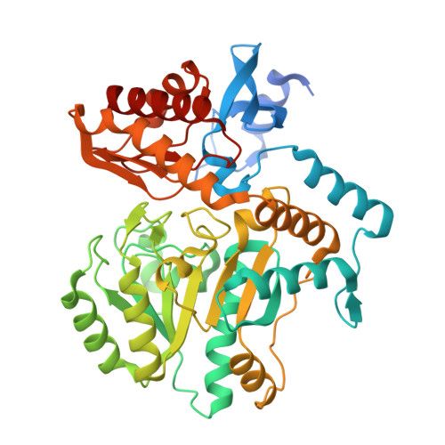
3TFT

Crystal structure of 7,8-diaminopelargonic acid synthase (BioA) from Mycobacterium tuberculosis, pre-reaction complex with a 3,6-dihydropyrid-2-one heterocycle inhibitor


Experimental Data Snapshot

  • Method: X-RAY DIFFRACTION
  • Resolution: 1.95 Å
  • R-Value Free: 0.197 
  • R-Value Work: 0.156 
  • R-Value Observed: 0.158 

wwPDB Validation   3D Report Full Report


Ligand Structure Quality Assessment 


This is version 1.2 of the entry. See complete history


Literature

Mechanism-based Inactivation by Aromatization of the Transaminase BioA Involved in Biotin Biosynthesis in Mycobaterium tuberculosis.

Shi, C.Geders, T.W.Park, S.W.Wilson, D.J.Boshoff, H.I.Abayomi, O.Barry, C.E.Schnappinger, D.Finzel, B.C.Aldrich, C.C.

(2011) J Am Chem Soc 133: 18194-18201

  • DOI: https://doi.org/10.1021/ja204036t
  • Primary Citation of Related Structures:  
    3TFT, 3TFU

  • PubMed Abstract: 

    BioA catalyzes the second step of biotin biosynthesis, and this enzyme represents a potential target to develop new antitubercular agents. Herein we report the design, synthesis, and biochemical characterization of a mechanism-based inhibitor (1) featuring a 3,6-dihydropyrid-2-one heterocycle that covalently modifies the pyridoxal 5'-phosphate (PLP) cofactor of BioA through aromatization. The structure of the PLP adduct was confirmed by MS/MS and X-ray crystallography at 1.94 Å resolution. Inactivation of BioA by 1 was time- and concentration-dependent and protected by substrate. We used a conditional knock-down mutant of M. tuberculosis to demonstrate the antitubercular activity of 1 correlated with BioA expression, and these results provide support for the designed mechanism of action.


  • Organizational Affiliation

    Academic Health Center, University of Minnesota, Minnesota 55455, United States.


Macromolecules
Find similar proteins by:  (by identity cutoff)  |  3D Structure
Entity ID: 1
MoleculeChains Sequence LengthOrganismDetailsImage
Adenosylmethionine-8-amino-7-oxononanoate aminotransferase
A, B
457Mycobacterium tuberculosisMutation(s): 0 
Gene Names: bioAMT1619MTCY336.35cRv1568
EC: 2.6.1.62
UniProt
Find proteins for P9WQ81 (Mycobacterium tuberculosis (strain ATCC 25618 / H37Rv))
Explore P9WQ81 
Go to UniProtKB:  P9WQ81
Entity Groups  
Sequence Clusters30% Identity50% Identity70% Identity90% Identity95% Identity100% Identity
UniProt GroupP9WQ81
Sequence Annotations
Expand
  • Reference Sequence
Experimental Data & Validation

Experimental Data

  • Method: X-RAY DIFFRACTION
  • Resolution: 1.95 Å
  • R-Value Free: 0.197 
  • R-Value Work: 0.156 
  • R-Value Observed: 0.158 
  • Space Group: P 21 21 21
Unit Cell:
Length ( Å )Angle ( ˚ )
a = 63.057α = 90
b = 66.385β = 90
c = 203.13γ = 90
Software Package:
Software NamePurpose
d*TREKdata scaling
d*TREKdata reduction
PHASERphasing
PHENIXrefinement
PDB_EXTRACTdata extraction
Blu-Icedata collection

Structure Validation

View Full Validation Report


Ligand Structure Quality Assessment 


Entry History 

Deposition Data

Revision History  (Full details and data files)

  • Version 1.0: 2011-10-19
    Type: Initial release
  • Version 1.1: 2011-11-23
    Changes: Database references
  • Version 1.2: 2023-09-13
    Changes: Data collection, Database references, Derived calculations, Refinement description