3TDV

Structure of the GDP complex of wild-type aminoglycoside 2'-phosphotransferase-IIIa


Experimental Data Snapshot

  • Method: X-RAY DIFFRACTION
  • Resolution: 2.20 Å
  • R-Value Free: 0.291 
  • R-Value Work: 0.204 
  • R-Value Observed: 0.209 

wwPDB Validation   3D Report Full Report


Ligand Structure Quality Assessment 


This is version 1.4 of the entry. See complete history


Literature

Aminoglycoside-2' phosphotransferase-IIIa (APH(2')-IIIa) prefers GTP over ATP: Structural templates for nucleotide recognition in the bacterial aminoglycoside-2' kinases.

Smith, C.A.Toth, M.Frase, H.Byrnes, L.J.Vakulenko, S.B.

(2012) J Biol Chem 287: 12893-12903

  • DOI: https://doi.org/10.1074/jbc.M112.341206
  • Primary Citation of Related Structures:  
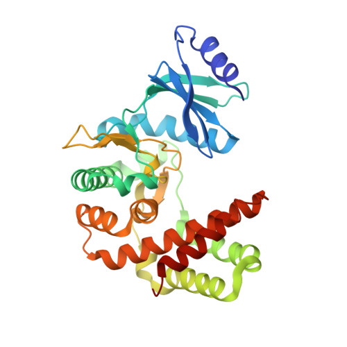
    3TDV, 3TDW

  • PubMed Abstract: 

    Contrary to the accepted dogma that ATP is the canonical phosphate donor in aminoglycoside kinases and protein kinases, it was recently demonstrated that all members of the bacterial aminoglycoside 2''-phosphotransferase IIIa (APH(2'')) aminoglycoside kinase family are unique in their ability to utilize GTP as a cofactor for antibiotic modification. Here we describe the structural determinants for GTP recognition in these enzymes. The crystal structure of the GTP-dependent APH(2'')-IIIa shows that although this enzyme has templates for both ATP and GTP binding superimposed on a single nucleotide specificity motif, access to the ATP-binding template is blocked by a bulky tyrosine residue. Substitution of this tyrosine by a smaller amino acid opens access to the ATP template. Similar GTP binding templates are conserved in other bacterial aminoglycoside kinases, whereas in the structurally related eukaryotic protein kinases this template is less conserved. The aminoglycoside kinases are important antibiotic resistance enzymes in bacteria, whose wide dissemination severely limits available therapeutic options, and the GTP binding templates could be exploited as new, previously unexplored targets for inhibitors of these clinically important enzymes.


  • Organizational Affiliation

    Stanford Synchrotron Radiation Lightsource, Stanford University, Menlo Park, California 94025, USA. csmith@slac.stanford.edu


Macromolecules
Find similar proteins by:  (by identity cutoff)  |  3D Structure
Entity ID: 1
MoleculeChains Sequence LengthOrganismDetailsImage
Gentamicin resistance protein
A, B
306Enterococcus gallinarumMutation(s): 0 
UniProt
Find proteins for P96762 (Enterococcus gallinarum)
Explore P96762 
Go to UniProtKB:  P96762
Entity Groups  
Sequence Clusters30% Identity50% Identity70% Identity90% Identity95% Identity100% Identity
UniProt GroupP96762
Sequence Annotations
Expand
  • Reference Sequence
Experimental Data & Validation

Experimental Data

  • Method: X-RAY DIFFRACTION
  • Resolution: 2.20 Å
  • R-Value Free: 0.291 
  • R-Value Work: 0.204 
  • R-Value Observed: 0.209 
  • Space Group: P 1
Unit Cell:
Length ( Å )Angle ( ˚ )
a = 41.68α = 89.94
b = 54.65β = 103.83
c = 77.16γ = 106.84
Software Package:
Software NamePurpose
Blu-Icedata collection
MOLREPphasing
PHENIXrefinement
XDSdata reduction
XSCALEdata scaling

Structure Validation

View Full Validation Report


Ligand Structure Quality Assessment 


Entry History 

Deposition Data

Revision History  (Full details and data files)

  • Version 1.0: 2012-03-07
    Type: Initial release
  • Version 1.1: 2012-03-14
    Changes: Database references
  • Version 1.2: 2012-05-02
    Changes: Database references
  • Version 1.3: 2020-02-26
    Changes: Data collection
  • Version 1.4: 2024-02-28
    Changes: Data collection, Database references, Derived calculations