3TCA

Crystal structure of the Ras-associating and pleckstrin-homology domains of RIAM


Experimental Data Snapshot

  • Method: X-RAY DIFFRACTION
  • Resolution: 2.35 Å
  • R-Value Free: 0.282 
  • R-Value Work: 0.234 
  • R-Value Observed: 0.236 

wwPDB Validation   3D Report Full Report


This is version 1.2 of the entry. See complete history


Literature

Rap1-interacting adapter molecule (RIAM) associates with the plasma membrane via a proximity detector.

Wynne, J.P.Wu, J.Su, W.Mor, A.Patsoukis, N.Boussiotis, V.A.Hubbard, S.R.Philips, M.R.

(2012) J Cell Biol 199: 317-329

  • DOI: https://doi.org/10.1083/jcb.201201157
  • Primary Citation of Related Structures:  
    3TCA

  • PubMed Abstract: 

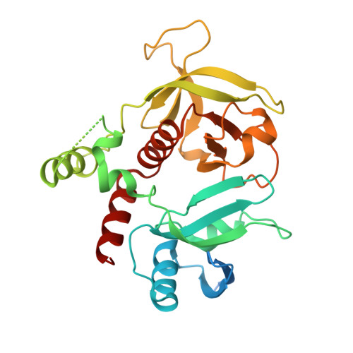
    Adaptive immunity depends on lymphocyte adhesion that is mediated by the integrin lymphocyte functional antigen 1 (LFA-1). The small guanosine triphosphatase Rap1 regulates LFA-1 adhesiveness through one of its effectors, Rap1-interacting adapter molecule (RIAM). We show that RIAM was recruited to the lymphocyte plasma membrane (PM) through its Ras association (RA) and pleckstrin homology (PH) domains, both of which were required for lymphocyte adhesion. The N terminus of RIAM inhibited membrane translocation. In vitro, the RA domain bound both Rap1 and H-Ras with equal but relatively low affinity, whereas in vivo only Rap1 was required for PM association. The PH domain bound phosphoinositol 4,5-bisphosphate (PI(4,5)P(2)) and was responsible for the spatial distribution of RIAM only at the PM of activated T cells. We determined the crystal structure of the RA and PH domains and found that, despite an intervening linker of 50 aa, the two domains were integrated into a single structural unit, which was critical for proper localization to the PM. Thus, the RA-PH domains of RIAM function as a proximity detector for activated Rap1 and PI(4,5)P(2).


  • Organizational Affiliation

    Cancer Institute, NYU School of Medicine, New York, NY 10016, USA.


Macromolecules
Find similar proteins by:  (by identity cutoff)  |  3D Structure
Entity ID: 1
MoleculeChains Sequence LengthOrganismDetailsImage
Amyloid beta A4 precursor protein-binding family B member 1-interacting protein
A, B
291Mus musculusMutation(s): 0 
Gene Names: Apbb1ipPrel1
UniProt & NIH Common Fund Data Resources
Find proteins for Q8R5A3 (Mus musculus)
Explore Q8R5A3 
Go to UniProtKB:  Q8R5A3
IMPC:  MGI:1861354
Entity Groups  
Sequence Clusters30% Identity50% Identity70% Identity90% Identity95% Identity100% Identity
UniProt GroupQ8R5A3
Sequence Annotations
Expand
  • Reference Sequence
Experimental Data & Validation

Experimental Data

  • Method: X-RAY DIFFRACTION
  • Resolution: 2.35 Å
  • R-Value Free: 0.282 
  • R-Value Work: 0.234 
  • R-Value Observed: 0.236 
  • Space Group: P 1 21 1
Unit Cell:
Length ( Å )Angle ( ˚ )
a = 46.86α = 90
b = 82.389β = 95.19
c = 83.404γ = 90
Software Package:
Software NamePurpose
ADSCdata collection
MOLREPphasing
REFMACrefinement
HKL-2000data reduction
HKL-2000data scaling

Structure Validation

View Full Validation Report


Entry History 

Deposition Data

Revision History  (Full details and data files)

  • Version 1.0: 2012-09-26
    Type: Initial release
  • Version 1.1: 2012-10-31
    Changes: Database references
  • Version 1.2: 2023-09-13
    Changes: Data collection, Database references, Refinement description