3T1W

Structure of the four-domain fragment Fn7B89 of oncofetal fibronectin


Experimental Data Snapshot

  • Method: X-RAY DIFFRACTION
  • Resolution: 2.40 Å
  • R-Value Free: 0.277 
  • R-Value Work: 0.228 
  • R-Value Observed: 0.230 

wwPDB Validation   3D Report Full Report


This is version 1.4 of the entry. See complete history


Literature

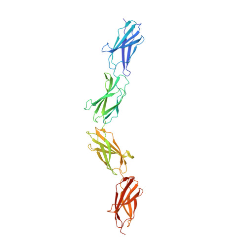
Extra-domain B in oncofetal fibronectin structurally promotes fibrillar head-to-tail dimerization of extracellular matrix protein.

Schiefner, A.Gebauer, M.Skerra, A.

(2012) J Biol Chem 287: 17578-17588

  • DOI: https://doi.org/10.1074/jbc.M111.303131
  • Primary Citation of Related Structures:  
    3T1W

  • PubMed Abstract: 

    The type III extra-domain B (ED-B) is specifically spliced into fibronectin (Fn) during embryogenesis and neoangiogenesis, including many cancers. The x-ray structure of the recombinant four-domain fragment Fn(III)7B89 reveals a tightly associated, extended head-to-tail dimer, which is stabilized via pair-wise shape and charge complementarity. A tendency toward ED-B-dependent dimer formation in solution was supported by size exclusion chromatography and analytical ultracentrifugation. When amending the model with the known three-dimensional structure of the Fn(III)10 domain, its RGD loop as well as the adhesion synergy region in Fn(III)9-10 become displayed on the same face of the dimer; this should allow simultaneous binding of at least two integrins and, thus, receptor clustering on the cell surface and intracellular signaling. Insertion of ED-B appears to stabilize overall head-to-tail dimerization of two separate Fn chains, which, together with alternating homodimer formation via disulfide bridges at the C-terminal Fn tail, should lead to the known macromolecular fibril formation.


  • Organizational Affiliation

    Munich Center for Integrated Protein Science (CIPS-M) and Lehrstuhl für Biologische Chemie, Technische Universität München, 85350 Freising-Weihenstephan, Germany.


Macromolecules
Find similar proteins by:  (by identity cutoff)  |  3D Structure
Entity ID: 1
MoleculeChains Sequence LengthOrganismDetailsImage
four-domain fibronectin fragment375Homo sapiensMutation(s): 0 
Gene Names: DKFZp686O12165NP_997647
UniProt & NIH Common Fund Data Resources
Find proteins for P02751 (Homo sapiens)
Explore P02751 
Go to UniProtKB:  P02751
PHAROS:  P02751
GTEx:  ENSG00000115414 
Entity Groups  
Sequence Clusters30% Identity50% Identity70% Identity90% Identity95% Identity100% Identity
UniProt GroupP02751
Sequence Annotations
Expand
  • Reference Sequence
Experimental Data & Validation

Experimental Data

  • Method: X-RAY DIFFRACTION
  • Resolution: 2.40 Å
  • R-Value Free: 0.277 
  • R-Value Work: 0.228 
  • R-Value Observed: 0.230 
  • Space Group: C 1 2 1
Unit Cell:
Length ( Å )Angle ( ˚ )
a = 211.98α = 90
b = 29.115β = 111.42
c = 82.241γ = 90
Software Package:
Software NamePurpose
MAR345data collection
PHASERphasing
REFMACrefinement
XDSdata reduction
XSCALEdata scaling

Structure Validation

View Full Validation Report


Entry History 

Deposition Data

Revision History  (Full details and data files)

  • Version 1.0: 2012-03-21
    Type: Initial release
  • Version 1.1: 2012-05-23
    Changes: Database references
  • Version 1.2: 2013-06-26
    Changes: Database references
  • Version 1.3: 2017-11-08
    Changes: Refinement description
  • Version 1.4: 2023-09-13
    Changes: Data collection, Database references, Refinement description