3SX2

Crystal structure of a putative 3-ketoacyl-(acyl-carrier-protein) reductase from Mycobacterium paratuberculosis in complex with NAD


Experimental Data Snapshot

  • Method: X-RAY DIFFRACTION
  • Resolution: 1.50 Å
  • R-Value Free: 0.159 
  • R-Value Work: 0.140 
  • R-Value Observed: 0.141 

wwPDB Validation   3D Report Full Report


Ligand Structure Quality Assessment 


This is version 1.4 of the entry. See complete history


Literature

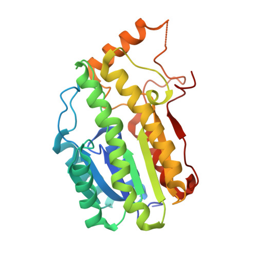
Mycofactocin-associated mycobacterial dehydrogenases with non-exchangeable NAD cofactors.

Haft, D.H.Pierce, P.G.Mayclin, S.J.Sullivan, A.Gardberg, A.S.Abendroth, J.Begley, D.W.Phan, I.Q.Staker, B.L.Myler, P.J.Marathias, V.M.Lorimer, D.D.Edwards, T.E.

(2017) Sci Rep 7: 41074-41074

  • DOI: https://doi.org/10.1038/srep41074
  • Primary Citation of Related Structures:  
    3OEC, 3PGX, 3PXX, 3S55, 3SX2, 3T7C, 3TSC, 4RGB, 5EJ2

  • PubMed Abstract: 

    During human infection, Mycobacterium tuberculosis (Mtb) survives the normally bacteriocidal phagosome of macrophages. Mtb and related species may be able to combat this harsh acidic environment which contains reactive oxygen species due to the mycobacterial genomes encoding a large number of dehydrogenases. Typically, dehydrogenase cofactor binding sites are open to solvent, which allows NAD/NADH exchange to support multiple turnover. Interestingly, mycobacterial short chain dehydrogenases/reductases (SDRs) within family TIGR03971 contain an insertion at the NAD binding site. Here we present crystal structures of 9 mycobacterial SDRs in which the insertion buries the NAD cofactor except for a small portion of the nicotinamide ring. Line broadening and STD-NMR experiments did not show NAD or NADH exchange on the NMR timescale. STD-NMR demonstrated binding of the potential substrate carveol, the potential product carvone, the inhibitor tricyclazol, and an external redox partner 2,6-dichloroindophenol (DCIP). Therefore, these SDRs appear to contain a non-exchangeable NAD cofactor and may rely on an external redox partner, rather than cofactor exchange, for multiple turnover. Incidentally, these genes always appear in conjunction with the mftA gene, which encodes the short peptide MftA, and with other genes proposed to convert MftA into the external redox partner mycofactocin.


  • Organizational Affiliation

    National Institutes of Health (NIH), Bethesda, MD 20892, USA.


Macromolecules
Find similar proteins by:  (by identity cutoff)  |  3D Structure
Entity ID: 1
MoleculeChains Sequence LengthOrganismDetailsImage
Putative 3-ketoacyl-(acyl-carrier-protein) reductase
A, B, C, D, E
A, B, C, D, E, F, G, H
278Mycobacterium avium subsp. paratuberculosis K-10Mutation(s): 0 
Gene Names: MAP_2861
UniProt
Find proteins for Q73W00 (Mycolicibacterium paratuberculosis (strain ATCC BAA-968 / K-10))
Explore Q73W00 
Go to UniProtKB:  Q73W00
Entity Groups  
Sequence Clusters30% Identity50% Identity70% Identity90% Identity95% Identity100% Identity
UniProt GroupQ73W00
Sequence Annotations
Expand
  • Reference Sequence
Small Molecules
Ligands 2 Unique
IDChains Name / Formula / InChI Key2D Diagram3D Interactions
NAD
Query on NAD

Download Ideal Coordinates CCD File 
I [auth A]
J [auth B]
L [auth C]
M [auth D]
O [auth E]
I [auth A],
J [auth B],
L [auth C],
M [auth D],
O [auth E],
Q [auth F],
S [auth G],
T [auth H]
NICOTINAMIDE-ADENINE-DINUCLEOTIDE
C21 H27 N7 O14 P2
BAWFJGJZGIEFAR-NNYOXOHSSA-N
MPD
Query on MPD

Download Ideal Coordinates CCD File 
K [auth B],
N [auth D],
P [auth E],
R [auth F],
U [auth H]
(4S)-2-METHYL-2,4-PENTANEDIOL
C6 H14 O2
SVTBMSDMJJWYQN-YFKPBYRVSA-N
Experimental Data & Validation

Experimental Data

  • Method: X-RAY DIFFRACTION
  • Resolution: 1.50 Å
  • R-Value Free: 0.159 
  • R-Value Work: 0.140 
  • R-Value Observed: 0.141 
  • Space Group: P 1
  • Diffraction Data: https://doi.org/10.18430/M33SX2
Unit Cell:
Length ( Å )Angle ( ˚ )
a = 64.46α = 97.01
b = 70.76β = 93.92
c = 125.68γ = 86.91
Software Package:
Software NamePurpose
BOSdata collection
PHASERphasing
REFMACrefinement
XDSdata reduction
XSCALEdata scaling

Structure Validation

View Full Validation Report


Ligand Structure Quality Assessment 


Entry History 

Deposition Data

Revision History  (Full details and data files)

  • Version 1.0: 2011-08-03
    Type: Initial release
  • Version 1.1: 2015-04-22
    Changes: Database references
  • Version 1.2: 2017-02-01
    Changes: Database references
  • Version 1.3: 2017-02-08
    Changes: Database references
  • Version 1.4: 2023-09-13
    Changes: Data collection, Database references, Derived calculations, Refinement description