3SGE

Crystal structure of mAb 17.2 in complex with R13 peptide


Experimental Data Snapshot

  • Method: X-RAY DIFFRACTION
  • Resolution: 1.89 Å
  • R-Value Free: 0.211 
  • R-Value Work: 0.179 
  • R-Value Observed: 0.181 

wwPDB Validation   3D Report Full Report


This is version 1.2 of the entry. See complete history


Literature

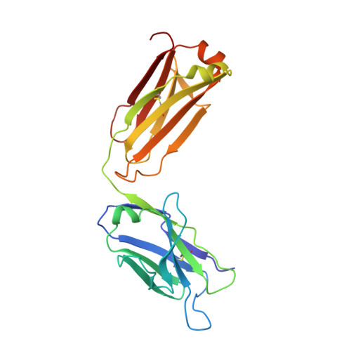
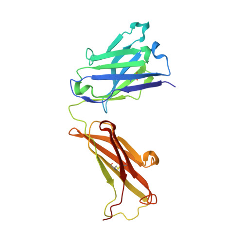
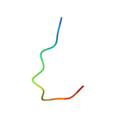
Crystal structure of the complex mAb 17.2 and the C-terminal region of Trypanosoma cruzi P2 Beta protein: implications in cross-reactivity

Pizarro, J.C.Boulot, G.Bentley, G.A.Gomez, K.A.Hoebeke, J.Hontebeyrie, M.Levin, M.J.Smulski, C.R.

(2011) PLoS Negl Trop Dis 5: e1375-e1375

  • DOI: https://doi.org/10.1371/journal.pntd.0001375
  • Primary Citation of Related Structures:  
    3SGD, 3SGE

  • PubMed Abstract: 

    Patients with Chronic Chagas' Heart Disease possess high levels of antibodies against the carboxyl-terminal end of the ribosomal P2ß protein of Trypanosoma cruzi (TcP2ß). These antibodies, as well as the murine monoclonal antibody (mAb) 17.2, recognize the last 13 amino acids of TcP2ß (called the R13 epitope: EEEDDDMGFGLFD) and are able to cross-react with, and stimulate, the ß1 adrenergic receptor (ß1-AR). Indeed, the mAb 17.2 was able to specifically detect human β1-AR, stably transfected into HEK cells, by flow cytometry and to induce repolarisation abnormalities and first degree atrioventricular conduction block after passive transfer to naïve mice. To study the structural basis of this cross-reactivity, we determined the crystal structure of the Fab region of the mAb 17.2 alone at 2.31 Å resolution and in complex with the R13 peptide at 1.89 Å resolution. We identified as key contact residues on R13 peptide Glu3, Asp6 and Phe9 as was previously shown by alanine scanning. Additionally, we generated a model of human β1-AR to elucidate the interaction with anti-R13 antibodies. These data provide an understanding of the molecular basis of cross-reactive antibodies induced by chronic infection with Trypanosoma cruzi.


  • Organizational Affiliation

    Unité d'Immunologie Structurale, Institut Pasteur, Paris, France.


Macromolecules
Find similar proteins by:  (by identity cutoff)  |  3D Structure
Entity ID: 1
MoleculeChains Sequence LengthOrganismDetailsImage
Light ChainA [auth L],
C [auth I]
219Mus musculusMutation(s): 0 
Entity Groups  
Sequence Clusters30% Identity50% Identity70% Identity90% Identity95% Identity100% Identity
Sequence Annotations
Expand
  • Reference Sequence
Find similar proteins by:  (by identity cutoff)  |  3D Structure
Entity ID: 2
MoleculeChains Sequence LengthOrganismDetailsImage
Heavy ChainB [auth H],
D [auth J]
217Mus musculusMutation(s): 0 
Entity Groups  
Sequence Clusters30% Identity50% Identity70% Identity90% Identity95% Identity100% Identity
Sequence Annotations
Expand
  • Reference Sequence

Find similar proteins by:  Sequence   |   3D Structure  

Entity ID: 3
MoleculeChains Sequence LengthOrganismDetailsImage
R13 peptideE [auth K],
F [auth M]
13N/AMutation(s): 0 
Entity Groups  
Sequence Clusters30% Identity50% Identity70% Identity90% Identity95% Identity100% Identity
Sequence Annotations
Expand
  • Reference Sequence
Experimental Data & Validation

Experimental Data

  • Method: X-RAY DIFFRACTION
  • Resolution: 1.89 Å
  • R-Value Free: 0.211 
  • R-Value Work: 0.179 
  • R-Value Observed: 0.181 
  • Space Group: P 1 21 1
Unit Cell:
Length ( Å )Angle ( ˚ )
a = 83α = 90
b = 68.21β = 98.37
c = 92.12γ = 90
Software Package:
Software NamePurpose
AMoREphasing
BUSTERrefinement
DENZOdata reduction
SCALEPACKdata scaling

Structure Validation

View Full Validation Report


Entry History 

Deposition Data

Revision History  (Full details and data files)

  • Version 1.0: 2011-11-09
    Type: Initial release
  • Version 1.1: 2013-01-23
    Changes: Database references
  • Version 1.2: 2023-09-13
    Changes: Data collection, Database references, Derived calculations, Refinement description