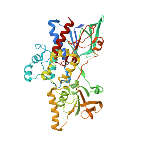
3S5W

Ornithine Hydroxylase (PvdA) from Pseudomonas aeruginosa


Experimental Data Snapshot

  • Method: X-RAY DIFFRACTION
  • Resolution: 1.90 Å
  • R-Value Free: 0.217 
  • R-Value Work: 0.192 
  • R-Value Observed: 0.193 

wwPDB Validation   3D Report Full Report


Ligand Structure Quality Assessment 


This is version 1.5 of the entry. See complete history


Literature

Two Structures of an N-Hydroxylating Flavoprotein Monooxygenase: ORNITHINE HYDROXYLASE FROM PSEUDOMONAS AERUGINOSA.

Olucha, J.Meneely, K.M.Chilton, A.S.Lamb, A.L.

(2011) J Biol Chem 286: 31789-31798

  • DOI: https://doi.org/10.1074/jbc.M111.265876
  • Primary Citation of Related Structures:  
    3S5W, 3S61

  • PubMed Abstract: 

    The ornithine hydroxylase from Pseudomonas aeruginosa (PvdA) catalyzes the FAD-dependent hydroxylation of the side chain amine of ornithine, which is subsequently formylated to generate the iron-chelating hydroxamates of the siderophore pyoverdin. PvdA belongs to the class B flavoprotein monooxygenases, which catalyze the oxidation of substrates using NADPH as the electron donor and molecular oxygen. Class B enzymes include the well studied flavin-containing monooxygenases and Baeyer-Villiger monooxygenases. The first two structures of a class B N-hydroxylating monooxygenase were determined with FAD in oxidized (1.9 Å resolution) and reduced (3.03 Å resolution) states. PvdA has the two expected Rossmann-like dinucleotide-binding domains for FAD and NADPH and also a substrate-binding domain, with the active site at the interface between the three domains. The structures have NADP(H) and (hydroxy)ornithine bound in a solvent-exposed active site, providing structural evidence for substrate and co-substrate specificity and the inability of PvdA to bind FAD tightly. Structural and biochemical evidence indicates that NADP(+) remains bound throughout the oxidative half-reaction, which is proposed to shelter the flavin intermediates from solvent and thereby prevent uncoupling of NADPH oxidation from hydroxylated product formation.


  • Organizational Affiliation

    Department of Molecular Biosciences, University of Kansas, Lawrence, Kansas 66045, USA.


Macromolecules
Find similar proteins by:  (by identity cutoff)  |  3D Structure
Entity ID: 1
MoleculeChains Sequence LengthOrganismDetailsImage
L-ornithine 5-monooxygenase
A, B
463Pseudomonas aeruginosaMutation(s): 0 
Gene Names: PA2386pvd-1pvdA
EC: 1.13.12
UniProt
Find proteins for Q51548 (Pseudomonas aeruginosa (strain ATCC 15692 / DSM 22644 / CIP 104116 / JCM 14847 / LMG 12228 / 1C / PRS 101 / PAO1))
Explore Q51548 
Go to UniProtKB:  Q51548
Entity Groups  
Sequence Clusters30% Identity50% Identity70% Identity90% Identity95% Identity100% Identity
UniProt GroupQ51548
Sequence Annotations
Expand
  • Reference Sequence
Small Molecules
Ligands 4 Unique
IDChains Name / Formula / InChI Key2D Diagram3D Interactions
FAD
Query on FAD

Download Ideal Coordinates CCD File 
C [auth A],
H [auth B]
FLAVIN-ADENINE DINUCLEOTIDE
C27 H33 N9 O15 P2
VWWQXMAJTJZDQX-UYBVJOGSSA-N
NAP
Query on NAP

Download Ideal Coordinates CCD File 
F [auth A],
I [auth B]
NADP NICOTINAMIDE-ADENINE-DINUCLEOTIDE PHOSPHATE
C21 H28 N7 O17 P3
XJLXINKUBYWONI-NNYOXOHSSA-N
ONH
Query on ONH

Download Ideal Coordinates CCD File 
E [auth A],
K [auth B]
N~5~-hydroxy-L-ornithine
C5 H12 N2 O3
OZMJDTPATROLQC-BYPYZUCNSA-N
PO4
Query on PO4

Download Ideal Coordinates CCD File 
D [auth A],
G [auth A],
J [auth B]
PHOSPHATE ION
O4 P
NBIIXXVUZAFLBC-UHFFFAOYSA-K
Experimental Data & Validation

Experimental Data

  • Method: X-RAY DIFFRACTION
  • Resolution: 1.90 Å
  • R-Value Free: 0.217 
  • R-Value Work: 0.192 
  • R-Value Observed: 0.193 
  • Space Group: I 41 2 2
Unit Cell:
Length ( Å )Angle ( ˚ )
a = 130.943α = 90
b = 130.943β = 90
c = 318.521γ = 90
Software Package:
Software NamePurpose
Blu-Icedata collection
PHASERphasing
REFMACrefinement
XDSdata reduction
XDSdata scaling

Structure Validation

View Full Validation Report


Ligand Structure Quality Assessment 


Entry History 

Deposition Data

Revision History  (Full details and data files)

  • Version 1.0: 2011-07-13
    Type: Initial release
  • Version 1.1: 2011-07-27
    Changes: Database references
  • Version 1.2: 2011-09-21
    Changes: Database references
  • Version 1.3: 2017-10-25
    Changes: Author supporting evidence
  • Version 1.4: 2024-02-28
    Changes: Data collection, Database references, Derived calculations
  • Version 1.5: 2024-04-03
    Changes: Refinement description