3S43

HIV-1 protease triple mutants V32I, I47V, V82I with antiviral drug amprenavir


Experimental Data Snapshot

  • Method: X-RAY DIFFRACTION
  • Resolution: 1.26 Å
  • R-Value Free: 0.200 
  • R-Value Work: 0.160 
  • R-Value Observed: 0.160 

wwPDB Validation   3D Report Full Report


Ligand Structure Quality Assessment 


This is version 1.1 of the entry. See complete history


Literature

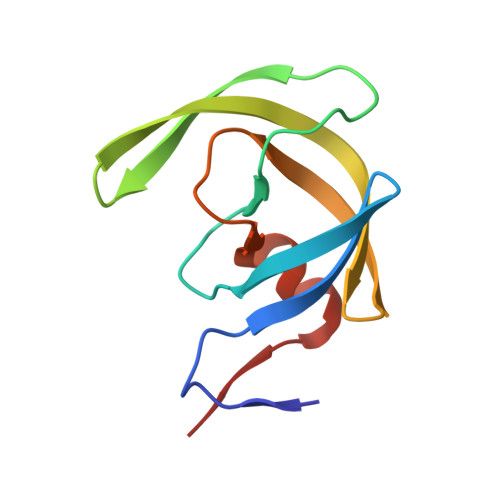
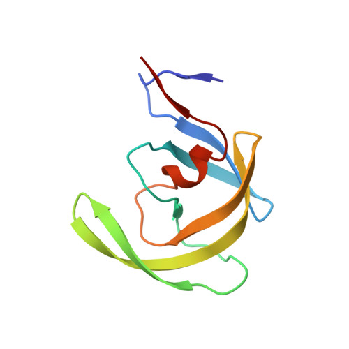
Critical differences in HIV-1 and HIV-2 protease specificity for clinical inhibitors.

Tie, Y.Wang, Y.F.Boross, P.I.Chiu, T.Y.Ghosh, A.K.Tozser, J.Louis, J.M.Harrison, R.W.Weber, I.T.

(2012) Protein Sci 21: 339-350

  • DOI: https://doi.org/10.1002/pro.2019
  • Primary Citation of Related Structures:  
    3S43, 3S45, 3S53, 3S54, 3S56

  • PubMed Abstract: 

    Clinical inhibitor amprenavir (APV) is less effective on HIV-2 protease (PR₂) than on HIV-1 protease (PR₁). We solved the crystal structure of PR₂ with APV at 1.5 Å resolution to identify structural changes associated with the lowered inhibition. Furthermore, we analyzed the PR₁ mutant (PR(1M) ) with substitutions V32I, I47V, and V82I that mimic the inhibitor binding site of PR₂. PR(1M) more closely resembled PR₂ than PR₁ in catalytic efficiency on four substrate peptides and inhibition by APV, whereas few differences were seen for two other substrates and inhibition by saquinavir (SQV) and darunavir (DRV). High resolution crystal structures of PR(1M) with APV, DRV, and SQV were compared with available PR₁ and PR₂ complexes. Val/Ile32 and Ile/Val47 showed compensating interactions with SQV in PR(1M) and PR₁, however, Ile82 interacted with a second SQV bound in an extension of the active site cavity of PR(1M). Residues 32 and 82 maintained similar interactions with DRV and APV in all the enzymes, whereas Val47 and Ile47 had opposing effects in the two subunits. Significantly diminished interactions were seen for the aniline of APV bound in PR₁ (M) and PR₂ relative to the strong hydrogen bonds observed in PR₁, consistent with 15- and 19-fold weaker inhibition, respectively. Overall, PR(1M) partially replicates the specificity of PR₂ and gives insight into drug resistant mutations at residues 32, 47, and 82. Moreover, this analysis provides a structural explanation for the weaker antiviral effects of APV on HIV-2.


  • Organizational Affiliation

    Department of Biology, Georgia State University, Atlanta, Georgia 30303, USA.


Macromolecules
Find similar proteins by:  (by identity cutoff)  |  3D Structure
Entity ID: 1
MoleculeChains Sequence LengthOrganismDetailsImage
Protease99Human immunodeficiency virus 1Mutation(s): 8 
Gene Names: pol
EC: 3.4.23.16
UniProt
Find proteins for Q7SSE3 (Human immunodeficiency virus 1)
Explore Q7SSE3 
Go to UniProtKB:  Q7SSE3
Entity Groups  
Sequence Clusters30% Identity50% Identity70% Identity90% Identity95% Identity100% Identity
UniProt GroupQ7SSE3
Sequence Annotations
Expand
  • Reference Sequence
Find similar proteins by:  (by identity cutoff)  |  3D Structure
Entity ID: 2
MoleculeChains Sequence LengthOrganismDetailsImage
Protease99Human immunodeficiency virus 1Mutation(s): 8 
Gene Names: pol
UniProt
Find proteins for Q7SSI0 (Human immunodeficiency virus 1)
Explore Q7SSI0 
Go to UniProtKB:  Q7SSI0
Entity Groups  
Sequence Clusters30% Identity50% Identity70% Identity90% Identity95% Identity100% Identity
UniProt GroupQ7SSI0
Sequence Annotations
Expand
  • Reference Sequence
Small Molecules
Ligands 3 Unique
IDChains Name / Formula / InChI Key2D Diagram3D Interactions
478
Query on 478

Download Ideal Coordinates CCD File 
C [auth A]{3-[(4-AMINO-BENZENESULFONYL)-ISOBUTYL-AMINO]-1-BENZYL-2-HYDROXY-PROPYL}-CARBAMIC ACID TETRAHYDRO-FURAN-3-YL ESTER
C25 H35 N3 O6 S
YMARZQAQMVYCKC-OEMFJLHTSA-N
IOD
Query on IOD

Download Ideal Coordinates CCD File 
D [auth A]
E [auth A]
F [auth A]
G [auth A]
H [auth A]
D [auth A],
E [auth A],
F [auth A],
G [auth A],
H [auth A],
I [auth A],
J [auth A],
K [auth A],
M [auth B],
N [auth B],
O [auth B],
P [auth B],
Q [auth B],
R [auth B],
S [auth B],
T [auth B],
U [auth B]
IODIDE ION
I
XMBWDFGMSWQBCA-UHFFFAOYSA-M
GOL
Query on GOL

Download Ideal Coordinates CCD File 
L [auth A],
V [auth B]
GLYCEROL
C3 H8 O3
PEDCQBHIVMGVHV-UHFFFAOYSA-N
Binding Affinity Annotations 
IDSourceBinding Affinity
478 BindingDB:  3S43 Ki: min: 7.00e-3, max: 57 (nM) from 9 assay(s)
Kd: min: 0.4, max: 0.59 (nM) from 2 assay(s)
Binding MOAD:  3S43 Ki: 2.53 (nM) from 1 assay(s)
PDBBind:  3S43 Ki: 2.53 (nM) from 1 assay(s)
Experimental Data & Validation

Experimental Data

  • Method: X-RAY DIFFRACTION
  • Resolution: 1.26 Å
  • R-Value Free: 0.200 
  • R-Value Work: 0.160 
  • R-Value Observed: 0.160 
  • Space Group: P 21 21 2
Unit Cell:
Length ( Å )Angle ( ˚ )
a = 58.374α = 90
b = 86.565β = 90
c = 46.306γ = 90
Software Package:
Software NamePurpose
HKL-2000data collection
AMoREphasing
SHELXL-97refinement
HKL-2000data reduction
HKL-2000data scaling

Structure Validation

View Full Validation Report


Ligand Structure Quality Assessment 


Entry History 

Deposition Data

Revision History  (Full details and data files)

  • Version 1.0: 2012-03-21
    Type: Initial release
  • Version 1.1: 2023-09-13
    Changes: Data collection, Database references, Derived calculations, Refinement description