3S3X

Structure of chicken acid-sensing ion channel 1 AT 3.0 A resolution in complex with psalmotoxin


Experimental Data Snapshot

  • Method: X-RAY DIFFRACTION
  • Resolution: 2.99 Å
  • R-Value Free: 0.249 
  • R-Value Work: 0.218 
  • R-Value Observed: 0.219 

wwPDB Validation   3D Report Full Report


Ligand Structure Quality Assessment 


This is version 1.5 of the entry. See complete history


Literature

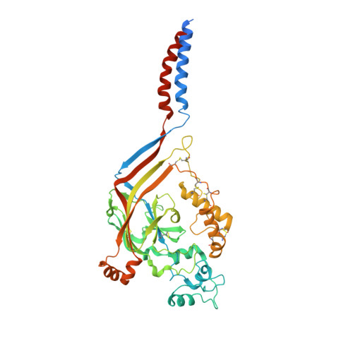
Structure of the Acid-sensing ion channel 1 in complex with the gating modifier Psalmotoxin 1.

Dawson, R.J.Benz, J.Stohler, P.Tetaz, T.Joseph, C.Huber, S.Schmid, G.Hugin, D.Pflimlin, P.Trube, G.Rudolph, M.G.Hennig, M.Ruf, A.

(2012) Nat Commun 3: 936-936

  • DOI: https://doi.org/10.1038/ncomms1917
  • Primary Citation of Related Structures:  
    3S3X

  • PubMed Abstract: 

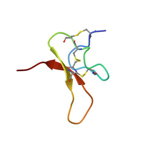
    Venom-derived peptide toxins can modify the gating characteristics of excitatory channels in neurons. How they bind and interfere with the flow of ions without directly blocking the ion permeation pathway remains elusive. Here we report the crystal structure of the trimeric chicken Acid-sensing ion channel 1 in complex with the highly selective gating modifier Psalmotoxin 1 at 3.0 Å resolution. The structure reveals the molecular interactions of three toxin molecules binding at the proton-sensitive acidic pockets of Acid-sensing ion channel 1 and electron density consistent with a cation trapped in the central vestibule above the ion pathway. A hydrophobic patch and a basic cluster are the key structural elements of Psalmotoxin 1 binding, locking two separate regulatory regions in their relative, desensitized-like arrangement. Our results provide a general concept for gating modifier toxin binding suggesting that both surface motifs are required to modify the gating characteristics of an ion channel.


  • Organizational Affiliation

    F. Hoffmann-La Roche AG, pRED, Pharma Research & Early Development, Discovery Technologies, Grenzacherstrasse 124, Basel CH4070, Switzerland. roger.dawson@roche.com


Macromolecules
Find similar proteins by:  (by identity cutoff)  |  3D Structure
Entity ID: 1
MoleculeChains Sequence LengthOrganismDetailsImage
Amiloride-sensitive cation channel 2, neuronal
A, B, C
459Gallus gallusMutation(s): 0 
Gene Names: ACCN2ASIC1
Membrane Entity: Yes 
UniProt
Find proteins for Q1XA76 (Gallus gallus)
Explore Q1XA76 
Go to UniProtKB:  Q1XA76
Entity Groups  
Sequence Clusters30% Identity50% Identity70% Identity90% Identity95% Identity100% Identity
UniProt GroupQ1XA76
Sequence Annotations
Expand
  • Reference Sequence
Find similar proteins by:  (by identity cutoff)  |  3D Structure
Entity ID: 2
MoleculeChains Sequence LengthOrganismDetailsImage
Psalmotoxin-1
D, E, F
37Psalmopoeus cambridgeiMutation(s): 0 
UniProt
Find proteins for P60514 (Psalmopoeus cambridgei)
Explore P60514 
Go to UniProtKB:  P60514
Entity Groups  
Sequence Clusters30% Identity50% Identity70% Identity90% Identity95% Identity100% Identity
UniProt GroupP60514
Sequence Annotations
Expand
  • Reference Sequence
Small Molecules
Ligands 4 Unique
IDChains Name / Formula / InChI Key2D Diagram3D Interactions
NAG
Query on NAG

Download Ideal Coordinates CCD File 
G [auth A]
H [auth A]
M [auth B]
N [auth B]
O [auth C]
G [auth A],
H [auth A],
M [auth B],
N [auth B],
O [auth C],
P [auth C]
2-acetamido-2-deoxy-beta-D-glucopyranose
C8 H15 N O6
OVRNDRQMDRJTHS-FMDGEEDCSA-N
SO4
Query on SO4

Download Ideal Coordinates CCD File 
L [auth A]SULFATE ION
O4 S
QAOWNCQODCNURD-UHFFFAOYSA-L
K
Query on K

Download Ideal Coordinates CCD File 
I [auth A],
J [auth A]
POTASSIUM ION
K
NPYPAHLBTDXSSS-UHFFFAOYSA-N
CL
Query on CL

Download Ideal Coordinates CCD File 
K [auth A]CHLORIDE ION
Cl
VEXZGXHMUGYJMC-UHFFFAOYSA-M
Experimental Data & Validation

Experimental Data

  • Method: X-RAY DIFFRACTION
  • Resolution: 2.99 Å
  • R-Value Free: 0.249 
  • R-Value Work: 0.218 
  • R-Value Observed: 0.219 
  • Space Group: C 1 2 1
Unit Cell:
Length ( Å )Angle ( ˚ )
a = 232.396α = 90
b = 109.442β = 119.81
c = 127.269γ = 90
Software Package:
Software NamePurpose
PHASERphasing
PHENIXrefinement
XDSdata reduction
SCALAdata scaling

Structure Validation

View Full Validation Report


Ligand Structure Quality Assessment 


Entry History 

Deposition Data

Revision History  (Full details and data files)

  • Version 1.0: 2012-05-23
    Type: Initial release
  • Version 1.1: 2012-06-06
    Changes: Structure summary
  • Version 1.2: 2012-07-18
    Changes: Database references
  • Version 1.3: 2017-11-08
    Changes: Refinement description
  • Version 1.4: 2020-07-29
    Type: Remediation
    Reason: Carbohydrate remediation
    Changes: Data collection, Database references, Derived calculations, Structure summary
  • Version 1.5: 2023-09-13
    Changes: Data collection, Database references, Refinement description, Structure summary