3RZC

Structure of the self-antigen iGb3 bound to mouse CD1d and in complex with the iNKT TCR


Experimental Data Snapshot

  • Method: X-RAY DIFFRACTION
  • Resolution: 2.80 Å
  • R-Value Free: 0.271 
  • R-Value Work: 0.236 
  • R-Value Observed: 0.237 

wwPDB Validation   3D Report Full Report


Ligand Structure Quality Assessment 


This is version 2.0 of the entry. See complete history


Literature

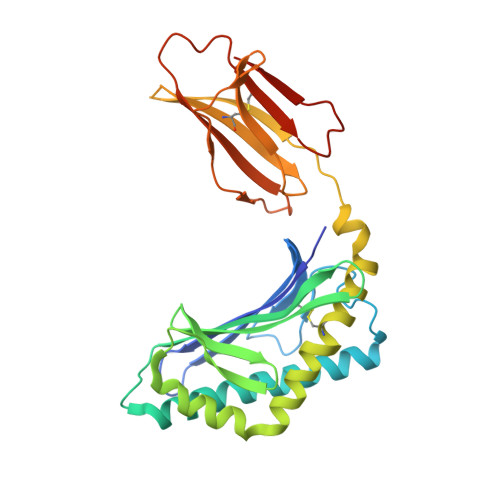
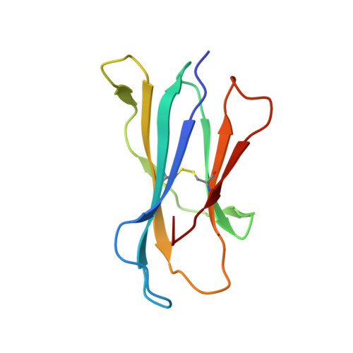
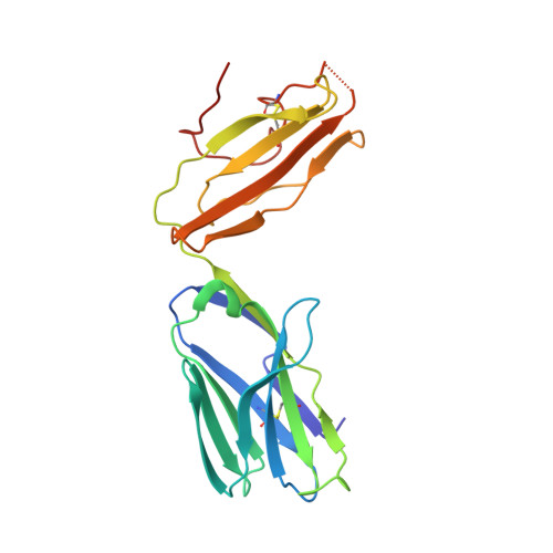
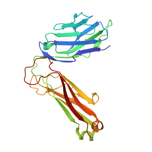
Cutting Edge: Structural Basis for the Recognition of {beta}-Linked Glycolipid Antigens by Invariant NKT Cells.

Yu, E.D.Girardi, E.Wang, J.Zajonc, D.M.

(2011) J Immunol 187: 2079-2083

  • DOI: https://doi.org/10.4049/jimmunol.1101636
  • Primary Citation of Related Structures:  
    3RZC

  • PubMed Abstract: 

    Invariant NKT (iNKT) cells expressing a semi-invariant Vα14 TCR recognize self and foreign lipid Ags when presented by the nonclassical MHCI homolog CD1d. Whereas the majority of known iNKT cell Ags are characterized by the presence of a single α-linked sugar, mammalian self Ags are β-linked glycosphingolipids, posing the interesting question of how the semi-invariant TCR can bind to such structurally distinct ligands. In this study, we show that the mouse iNKT TCR recognizes the complex β-linked Ag isoglobotrihexosylceramide (iGb3; Galα1-3-Galβ1-4-Glcβ1-1Cer) by forcing the proximal β-linked sugar of the trisaccharide head group to adopt the typical binding orientation of α-linked glycolipids. The squashed iGb3 orientation is stabilized by several interactions between the trisaccharide and CD1d residues. Finally, the formation of novel contacts between the proximal and second sugar of iGb3 and CDR2α residues of the TCR suggests an expanded recognition logic that can possibly distinguish foreign Ags from self Ags.


  • Organizational Affiliation

    Division of Cell Biology, La Jolla Institute for Allergy and Immunology, La Jolla, CA 92037, USA.


Macromolecules
Find similar proteins by:  (by identity cutoff)  |  3D Structure
Entity ID: 1
MoleculeChains Sequence LengthOrganismDetailsImage
Antigen-presenting glycoprotein CD1d1285Mus musculusMutation(s): 0 
Gene Names: Cd1.1CD1dCd1d1
UniProt & NIH Common Fund Data Resources
Find proteins for P11609 (Mus musculus)
Explore P11609 
Go to UniProtKB:  P11609
IMPC:  MGI:107674
Entity Groups  
Sequence Clusters30% Identity50% Identity70% Identity90% Identity95% Identity100% Identity
UniProt GroupP11609
Sequence Annotations
Expand
  • Reference Sequence
Find similar proteins by:  (by identity cutoff)  |  3D Structure
Entity ID: 2
MoleculeChains Sequence LengthOrganismDetailsImage
Beta-2-microglobulin99Mus musculusMutation(s): 1 
Gene Names: B2mbeta-2-microglobulin
UniProt & NIH Common Fund Data Resources
Find proteins for P01887 (Mus musculus)
Explore P01887 
Go to UniProtKB:  P01887
IMPC:  MGI:88127
Entity Groups  
Sequence Clusters30% Identity50% Identity70% Identity90% Identity95% Identity100% Identity
UniProt GroupP01887
Sequence Annotations
Expand
  • Reference Sequence
Find similar proteins by:  (by identity cutoff)  |  3D Structure
Entity ID: 3
MoleculeChains Sequence LengthOrganismDetailsImage
Valpha14209Mus musculusHomo sapiens
This entity is chimeric
Mutation(s): 0 
Gene Names: Valpha14
Entity Groups  
Sequence Clusters30% Identity50% Identity70% Identity90% Identity95% Identity100% Identity
Sequence Annotations
Expand
  • Reference Sequence
Find similar proteins by:  (by identity cutoff)  |  3D Structure
Entity ID: 4
MoleculeChains Sequence LengthOrganismDetailsImage
Vbeta8.2241Mus musculusHomo sapiens
This entity is chimeric
Mutation(s): 0 
Gene Names: Vbeta8.2
Entity Groups  
Sequence Clusters30% Identity50% Identity70% Identity90% Identity95% Identity100% Identity
Sequence Annotations
Expand
  • Reference Sequence
Oligosaccharides

Help

Entity ID: 5
MoleculeChains Length2D Diagram Glycosylation3D Interactions
2-acetamido-2-deoxy-beta-D-glucopyranose-(1-4)-2-acetamido-2-deoxy-beta-D-glucopyranose
E, F
2N-Glycosylation
Glycosylation Resources
GlyTouCan:  G42666HT
GlyCosmos:  G42666HT
GlyGen:  G42666HT
Entity ID: 6
MoleculeChains Length2D Diagram Glycosylation3D Interactions
alpha-D-mannopyranose-(1-4)-2-acetamido-2-deoxy-beta-D-glucopyranose-(1-4)-[beta-L-fucopyranose-(1-6)]2-acetamido-2-deoxy-beta-D-glucopyranose
G
4N-Glycosylation
Glycosylation Resources
GlyTouCan:  G70885YM
GlyCosmos:  G70885YM
GlyGen:  G70885YM
Small Molecules
Ligands 1 Unique
IDChains Name / Formula / InChI Key2D Diagram3D Interactions
LGN
Query on LGN

Download Ideal Coordinates CCD File 
H [auth A]N-[(2S,3R,4E)-1-{[alpha-D-galactopyranosyl-(1->3)-beta-D-galactopyranosyl-(1->4)-beta-D-glucopyranosyl]oxy}-3-hydroxyoctadec-4-en-2-yl]hexacosanamide
C62 H117 N O18
JMENXJYBCQFIRK-KRJDXUSZSA-N
Experimental Data & Validation

Experimental Data

  • Method: X-RAY DIFFRACTION
  • Resolution: 2.80 Å
  • R-Value Free: 0.271 
  • R-Value Work: 0.236 
  • R-Value Observed: 0.237 
  • Space Group: C 2 2 21
Unit Cell:
Length ( Å )Angle ( ˚ )
a = 79.103α = 90
b = 191.346β = 90
c = 151.032γ = 90
Software Package:
Software NamePurpose
HKL-2000data collection
MOLREPphasing
REFMACrefinement
HKL-2000data reduction
HKL-2000data scaling

Structure Validation

View Full Validation Report


Ligand Structure Quality Assessment 


Entry History 

Deposition Data

Revision History  (Full details and data files)

  • Version 1.0: 2011-08-24
    Type: Initial release
  • Version 1.1: 2011-09-07
    Changes: Database references
  • Version 1.2: 2013-09-18
    Changes: Database references
  • Version 2.0: 2020-07-29
    Type: Remediation
    Reason: Carbohydrate remediation
    Changes: Atomic model, Data collection, Database references, Derived calculations, Structure summary