


Crystal structure of putative polysaccharide deacetylase from Mycobacterium smegmatis
Michalska, K., Tesar, C., Bearden, J., Joachimiak, A.To be published.
Experimental Data Snapshot
wwPDB Validation 3D Report Full Report

Entity ID: 1 | |||||
|---|---|---|---|---|---|
| Molecule | Chains | Sequence Length | Organism | Details | Image |
| Polysaccharide deacetylase | 300 | Mycolicibacterium smegmatis MC2 155 | Mutation(s): 0 Gene Names: MSMEG_4373 | 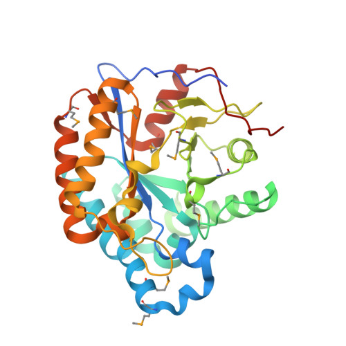 | |
UniProt | |||||
Find proteins for A0R0G0 (Mycolicibacterium smegmatis (strain ATCC 700084 / mc(2)155)) Explore A0R0G0 Go to UniProtKB: A0R0G0 | |||||
Entity Groups | |||||
| Sequence Clusters | 30% Identity50% Identity70% Identity90% Identity95% Identity100% Identity | ||||
| UniProt Group | A0R0G0 | ||||
Sequence AnnotationsExpand | |||||
| |||||
| Ligands 2 Unique | |||||
|---|---|---|---|---|---|
| ID | Chains | Name / Formula / InChI Key | 2D Diagram | 3D Interactions | |
| ZN Query on ZN | F [auth A], H [auth B], J [auth C], L [auth D] | ZINC ION Zn PTFCDOFLOPIGGS-UHFFFAOYSA-N |  | ||
| CL Query on CL | E [auth A], G [auth B], I [auth C], K [auth D] | CHLORIDE ION Cl VEXZGXHMUGYJMC-UHFFFAOYSA-M |  | ||
| Modified Residues 1 Unique | |||||
|---|---|---|---|---|---|
| ID | Chains | Type | Formula | 2D Diagram | Parent |
| MSE Query on MSE | A, B, C, D | L-PEPTIDE LINKING | C5 H11 N O2 Se |  | MET |
| Length ( Å ) | Angle ( ˚ ) |
|---|---|
| a = 80.212 | α = 90 |
| b = 59.592 | β = 95.35 |
| c = 130.387 | γ = 90 |
| Software Name | Purpose |
|---|---|
| SBC-Collect | data collection |
| Auto-Rickshaw | phasing |
| SHELXD | phasing |
| SHELXE | model building |
| RESOLVE | model building |
| DM | model building |
| ARP/wARP | model building |
| BUSTER | refinement |
| HKL-3000 | data reduction |
| HKL-3000 | data scaling |
| RESOLVE | phasing |
| DM | phasing |
RCSB PDB (citation) is hosted by
RCSB PDB is a member of the

