3RIX

1.7A resolution structure of a firefly luciferase-Aspulvinone J inhibitor complex


Experimental Data Snapshot

  • Method: X-RAY DIFFRACTION
  • Resolution: 1.70 Å
  • R-Value Free: 0.218 
  • R-Value Work: 0.190 
  • R-Value Observed: 0.191 

wwPDB Validation   3D Report Full Report


Ligand Structure Quality Assessment 


This is version 1.4 of the entry. See complete history


Literature

Titration-based screening for evaluation of natural product extracts: identification of an aspulvinone family of luciferase inhibitors.

Cruz, P.G.Auld, D.S.Schultz, P.J.Lovell, S.Battaile, K.P.MacArthur, R.Shen, M.Tamayo-Castillo, G.Inglese, J.Sherman, D.H.

(2011) Chem Biol 18: 1442-1452

  • DOI: https://doi.org/10.1016/j.chembiol.2011.08.011
  • Primary Citation of Related Structures:  
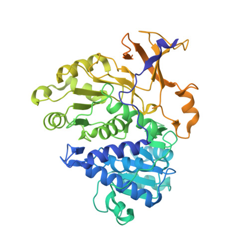
    3RIX

  • PubMed Abstract: 

    The chemical diversity of nature has tremendous potential for the discovery of molecular probes and medicinal agents. However, sensitivity of HTS assays to interfering components of crude extracts derived from plants, and macro- and microorganisms has curtailed their use in lead discovery. Here, we describe a process for leveraging the concentration-response curves obtained from quantitative HTS to improve the initial selection of "actives" from a library of partially fractionated natural product extracts derived from marine actinomycetes and fungi. By using pharmacological activity, the first-pass CRC paradigm improves the probability that labor-intensive subsequent steps of reculturing, extraction, and bioassay-guided isolation of active component(s) target the most promising strains and growth conditions. We illustrate how this process identified a family of fungal metabolites as potent inhibitors of firefly luciferase, subsequently resolved in molecular detail by X-ray crystallography.


  • Organizational Affiliation

    Life Sciences Institute, University of Michigan, Ann Arbor, MI 48109-2216, USA.


Macromolecules
Find similar proteins by:  (by identity cutoff)  |  3D Structure
Entity ID: 1
MoleculeChains Sequence LengthOrganismDetailsImage
Luciferin 4-monooxygenase550Photinus pyralisMutation(s): 0 
EC: 1.13.12.7
UniProt
Find proteins for P08659 (Photinus pyralis)
Explore P08659 
Go to UniProtKB:  P08659
Entity Groups  
Sequence Clusters30% Identity50% Identity70% Identity90% Identity95% Identity100% Identity
UniProt GroupP08659
Sequence Annotations
Expand
  • Reference Sequence
Small Molecules
Ligands 1 Unique
IDChains Name / Formula / InChI Key2D Diagram3D Interactions
923
Query on 923

Download Ideal Coordinates CCD File 
B [auth A](5Z)-4-hydroxy-3-[(2R)-2-(2-hydroxypropan-2-yl)-2,3-dihydro-1-benzofuran-5-yl]-5-{[(2R)-2-(2-hydroxypropan-2-yl)-2,3-dihydro-1-benzofuran-5-yl]methylidene}furan-2(5H)-one
C27 H28 O7
YPXKKOMPEGAWMY-JQOCIHOLSA-N
Experimental Data & Validation

Experimental Data

  • Method: X-RAY DIFFRACTION
  • Resolution: 1.70 Å
  • R-Value Free: 0.218 
  • R-Value Work: 0.190 
  • R-Value Observed: 0.191 
  • Space Group: P 41
Unit Cell:
Length ( Å )Angle ( ˚ )
a = 83.738α = 90
b = 83.738β = 90
c = 96.913γ = 90
Software Package:
Software NamePurpose
SCALAdata scaling
PHENIXrefinement
PDB_EXTRACTdata extraction
JDirectordata collection
XDSdata reduction

Structure Validation

View Full Validation Report


Ligand Structure Quality Assessment 


Entry History 

Deposition Data

Revision History  (Full details and data files)

  • Version 1.0: 2011-12-07
    Type: Initial release
  • Version 1.1: 2012-03-28
    Changes: Database references
  • Version 1.2: 2012-12-05
    Changes: Non-polymer description
  • Version 1.3: 2017-11-08
    Changes: Refinement description
  • Version 1.4: 2023-09-13
    Changes: Data collection, Database references, Derived calculations, Refinement description