3RGH

Structure of filamin A immunoglobulin-like repeat 10 from Homo sapiens


Experimental Data Snapshot

  • Method: X-RAY DIFFRACTION
  • Resolution: 2.44 Å
  • R-Value Free: 0.279 
  • R-Value Work: 0.229 
  • R-Value Observed: 0.234 

wwPDB Validation   3D Report Full Report


This is version 1.2 of the entry. See complete history


Literature

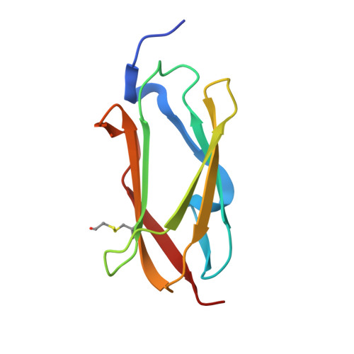
Structure of filamin A immunoglobulin-like repeat 10 from Homo sapiens.

Page, R.C.Clark, J.G.Misra, S.

(2011) Acta Crystallogr Sect F Struct Biol Cryst Commun 67: 871-876

  • DOI: https://doi.org/10.1107/S1744309111024249
  • Primary Citation of Related Structures:  
    3RGH

  • PubMed Abstract: 

    Filamin A (FlnA) plays a critical role in cytoskeletal organization, cell motility and cellular signaling. FlnA utilizes different binding sites on a series of 24 immunoglobulin-like domains (Ig repeats) to interact with diverse cytosolic proteins and with cytoplasmic portions of membrane proteins. Mutations in a specific domain, Ig10 (FlnA-Ig10), are correlated with two severe forms of the otopalatodigital syndrome spectrum disorders Melnick-Needles syndrome and frontometaphyseal dysplasia. The crystal structure of FlnA-Ig10 determined at 2.44 Å resolution provides insight into the perturbations caused by these mutations.


  • Organizational Affiliation

    Department of Molecular Cardiology, Lerner Research Institute, Cleveland Clinic, Cleveland, OH 44195, USA. pager2@ccf.org


Macromolecules
Find similar proteins by:  (by identity cutoff)  |  3D Structure
Entity ID: 1
MoleculeChains Sequence LengthOrganismDetailsImage
Filamin-A
A, B
100Homo sapiensMutation(s): 0 
Gene Names: FLNFLN1FLNA
UniProt & NIH Common Fund Data Resources
Find proteins for P21333 (Homo sapiens)
Explore P21333 
Go to UniProtKB:  P21333
PHAROS:  P21333
GTEx:  ENSG00000196924 
Entity Groups  
Sequence Clusters30% Identity50% Identity70% Identity90% Identity95% Identity100% Identity
UniProt GroupP21333
Sequence Annotations
Expand
  • Reference Sequence
Small Molecules
Modified Residues  1 Unique
IDChains TypeFormula2D DiagramParent
CME
Query on CME
A, B
L-PEPTIDE LINKINGC5 H11 N O3 S2CYS
Experimental Data & Validation

Experimental Data

  • Method: X-RAY DIFFRACTION
  • Resolution: 2.44 Å
  • R-Value Free: 0.279 
  • R-Value Work: 0.229 
  • R-Value Observed: 0.234 
  • Space Group: P 21 21 21
Unit Cell:
Length ( Å )Angle ( ˚ )
a = 36.453α = 90
b = 50.549β = 90
c = 107.172γ = 90
Software Package:
Software NamePurpose
StructureStudiodata collection
PHENIXmodel building
PHENIXrefinement
d*TREKdata reduction
d*TREKdata scaling
PHENIXphasing

Structure Validation

View Full Validation Report


Entry History 

Deposition Data

Revision History  (Full details and data files)

  • Version 1.0: 2011-08-03
    Type: Initial release
  • Version 1.1: 2011-08-17
    Changes: Database references
  • Version 1.2: 2024-04-03
    Changes: Data collection, Database references, Derived calculations, Refinement description