Structure and Mechanism of the Saposin-like Domain of a Plant Aspartic Protease.
Bryksa, B.C., Bhaumik, P., Magracheva, E., De Moura, D.C., Kurylowicz, M., Zdanov, A., Dutcher, J.R., Wlodawer, A., Yada, R.Y.(2011) J Biol Chem 286: 28265-28275
- PubMed: 21676875
- DOI: https://doi.org/10.1074/jbc.M111.252619
- Primary Citation of Related Structures:
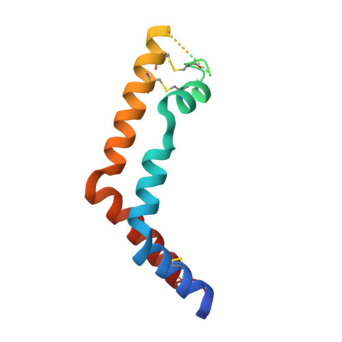
3RFI - PubMed Abstract:
Many plant aspartic proteases contain an additional sequence of ~100 amino acids termed the plant-specific insert, which is involved in host defense and vacuolar targeting. Similar to all saposin-like proteins, the plant-specific insert functions via protein-membrane interactions; however, the structural basis for such interactions has not been studied, and the nature of plant-specific insert-mediated membrane disruption has not been characterized. In the present study, the crystal structure of the saposin-like domain of potato aspartic protease was resolved at a resolution of 1.9 Å, revealing an open V-shaped configuration similar to the open structure of human saposin C. Notably, vesicle disruption activity followed Michaelis-Menten-like kinetics, a finding not previously reported for saposin-like proteins including plant-specific inserts. Circular dichroism data suggested that secondary structure was pH-dependent in a fashion similar to influenza A hemagglutinin fusion peptide. Membrane effects characterized by atomic force microscopy and light scattering indicated bilayer solubilization as well as fusogenic activity. Taken together, the present study is the first report to elucidate the membrane interaction mechanism of plant saposin-like domains whereby pH-dependent membrane interactions resulted in bilayer fusogenic activity that probably arose from a viral type pH-dependent helix-kink-helix motif at the plant-specific insert N terminus.
Organizational Affiliation:
Department of Food Science, University of Guelph, Guelph, Ontario N1G 2W1, Canada.