3RE2

Crystal structure of menin reveals the binding site for Mixed Lineage Leukemia (MLL) protein


Experimental Data Snapshot

  • Method: X-RAY DIFFRACTION
  • Resolution: 1.95 Å
  • R-Value Free: 0.211 
  • R-Value Work: 0.161 
  • R-Value Observed: 0.163 

wwPDB Validation   3D Report Full Report


This is version 1.2 of the entry. See complete history


Literature

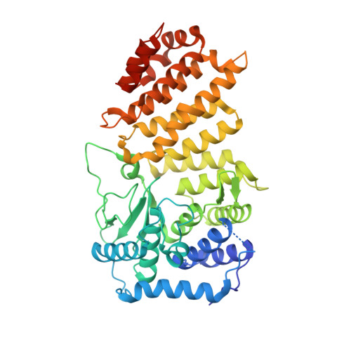
Crystal Structure of Menin Reveals Binding Site for Mixed Lineage Leukemia (MLL) Protein.

Murai, M.J.Chruszcz, M.Reddy, G.Grembecka, J.Cierpicki, T.

(2011) J Biol Chem 286: 31742-31748

  • DOI: https://doi.org/10.1074/jbc.M111.258186
  • Primary Citation of Related Structures:  
    3RE2

  • PubMed Abstract: 

    Menin is a tumor suppressor protein that is encoded by the MEN1 (multiple endocrine neoplasia 1) gene and controls cell growth in endocrine tissues. Importantly, menin also serves as a critical oncogenic cofactor of MLL (mixed lineage leukemia) fusion proteins in acute leukemias. Direct association of menin with MLL fusion proteins is required for MLL fusion protein-mediated leukemogenesis in vivo, and this interaction has been validated as a new potential therapeutic target for development of novel anti-leukemia agents. Here, we report the first crystal structure of menin homolog from Nematostella vectensis. Due to a very high sequence similarity, the Nematostella menin is a close homolog of human menin, and these two proteins likely have very similar structures. Menin is predominantly an α-helical protein with the protein core comprising three tetratricopeptide motifs that are flanked by two α-helical bundles and covered by a β-sheet motif. A very interesting feature of menin structure is the presence of a large central cavity that is highly conserved between Nematostella and human menin. By employing site-directed mutagenesis, we have demonstrated that this cavity constitutes the binding site for MLL. Our data provide a structural basis for understanding the role of menin as a tumor suppressor protein and as an oncogenic co-factor of MLL fusion proteins. It also provides essential structural information for development of inhibitors targeting the menin-MLL interaction as a novel therapeutic strategy in MLL-related leukemias.


  • Organizational Affiliation

    Department of Pathology, University of Michigan, Ann Arbor, Michigan 48109, USA.


Macromolecules
Find similar proteins by:  (by identity cutoff)  |  3D Structure
Entity ID: 1
MoleculeChains Sequence LengthOrganismDetailsImage
Predicted protein472Nematostella vectensisMutation(s): 0 
Gene Names: v1g241841
UniProt
Find proteins for A7RZU9 (Nematostella vectensis)
Explore A7RZU9 
Go to UniProtKB:  A7RZU9
Entity Groups  
Sequence Clusters30% Identity50% Identity70% Identity90% Identity95% Identity100% Identity
UniProt GroupA7RZU9
Sequence Annotations
Expand
  • Reference Sequence
Small Molecules
Ligands 1 Unique
IDChains Name / Formula / InChI Key2D Diagram3D Interactions
GOL
Query on GOL

Download Ideal Coordinates CCD File 
B [auth A]GLYCEROL
C3 H8 O3
PEDCQBHIVMGVHV-UHFFFAOYSA-N
Experimental Data & Validation

Experimental Data

  • Method: X-RAY DIFFRACTION
  • Resolution: 1.95 Å
  • R-Value Free: 0.211 
  • R-Value Work: 0.161 
  • R-Value Observed: 0.163 
  • Space Group: P 43 21 2
Unit Cell:
Length ( Å )Angle ( ˚ )
a = 89.881α = 90
b = 89.881β = 90
c = 115.601γ = 90
Software Package:
Software NamePurpose
HKL-3000data collection
MD2data collection
HKL-3000phasing
SHELXDphasing
SHELXEmodel building
MLPHAREphasing
DMmodel building
RESOLVEmodel building
ARP/wARPmodel building
CCP4model building
REFMACrefinement
Cootmodel building
HKL-2000data reduction
HKL-2000data scaling
DMphasing
RESOLVEphasing
CCP4phasing

Structure Validation

View Full Validation Report


Entry History 

Deposition Data

Revision History  (Full details and data files)

  • Version 1.0: 2011-07-13
    Type: Initial release
  • Version 1.1: 2011-09-21
    Changes: Database references
  • Version 1.2: 2024-02-21
    Changes: Data collection, Database references, Derived calculations