3QS1

Crystal structure of KNI-10006 complex of Plasmepsin I (PMI) from Plasmodium falciparum


Experimental Data Snapshot

  • Method: X-RAY DIFFRACTION
  • Resolution: 3.10 Å
  • R-Value Free: 0.300 
  • R-Value Work: 0.211 
  • R-Value Observed: 0.215 

wwPDB Validation   3D Report Full Report


Ligand Structure Quality Assessment 


This is version 1.2 of the entry. See complete history


Literature

Crystal structures of the free and inhibited forms of plasmepsin I (PMI) from Plasmodium falciparum.

Bhaumik, P.Horimoto, Y.Xiao, H.Miura, T.Hidaka, K.Kiso, Y.Wlodawer, A.Yada, R.Y.Gustchina, A.

(2011) J Struct Biol 175: 73-84

  • DOI: https://doi.org/10.1016/j.jsb.2011.04.009
  • Primary Citation of Related Structures:  
    3QRV, 3QS1

  • PubMed Abstract: 

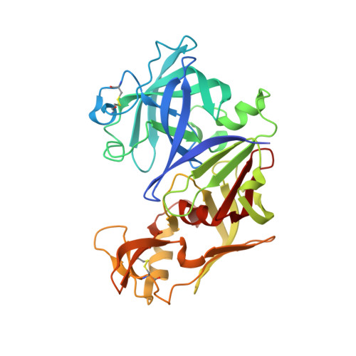
    Plasmepsin I (PMI) is one of the four vacuolar pepsin-like proteases responsible for hemoglobin degradation by the malarial parasite Plasmodium falciparum, and the only one with no crystal structure reported to date. Due to substantial functional redundancy of these enzymes, lack of inhibition of even a single plasmepsin can defeat efforts in creating effective antiparasitic agents. We have now solved crystal structures of the recombinant PMI as apoenzyme and in complex with the potent peptidic inhibitor, KNI-10006, at the resolution of 2.4 and 3.1Å, respectively. The apoenzyme crystallized in the orthorhombic space group P2(1)2(1)2(1) with two molecules in the asymmetric unit and the structure has been refined to the final R-factor of 20.7%. The KNI-10006 bound enzyme crystallized in the tetragonal space group P4(3) with four molecules in the asymmetric unit and the structure has been refined to the final R-factor of 21.1%. In the PMI-KNI-10006 complex, the inhibitors were bound identically to all four enzyme molecules, with the opposite directionality of the main chain of KNI-10006 relative to the direction of the enzyme substrates. Such a mode of binding of inhibitors containing an allophenylnorstatine-dimethylthioproline insert in the P1-P1' positions, previously reported in a complex with PMIV, demonstrates the importance of satisfying the requirements for the proper positioning of the functional groups in the mechanism-based inhibitors towards the catalytic machinery of aspartic proteases, as opposed to binding driven solely by the specificity of the individual enzymes. A comparison of the structure of the PMI-KNI-10006 complex with the structures of other vacuolar plasmepsins identified the important differences between them and may help in the design of specific inhibitors targeting the individual enzymes.


  • Organizational Affiliation

    Protein Structure Section, Macromolecular Crystallography Laboratory, National Cancer Institute, Frederick, MD 21702, USA.


Macromolecules
Find similar proteins by:  (by identity cutoff)  |  3D Structure
Entity ID: 1
MoleculeChains Sequence LengthOrganismDetailsImage
Plasmepsin-1
A, B, C, D
336Plasmodium falciparumMutation(s): 0 
Gene Names: PMI
EC: 3.4.23.38
UniProt
Find proteins for P39898 (Plasmodium falciparum (isolate HB3))
Explore P39898 
Go to UniProtKB:  P39898
Entity Groups  
Sequence Clusters30% Identity50% Identity70% Identity90% Identity95% Identity100% Identity
UniProt GroupP39898
Sequence Annotations
Expand
  • Reference Sequence
Small Molecules
Ligands 2 Unique
IDChains Name / Formula / InChI Key2D Diagram3D Interactions
006
Query on 006

Download Ideal Coordinates CCD File 
E [auth A],
I [auth B],
L [auth C],
P [auth D]
(4R)-3-[(2S,3S)-3-{[(2,6-dimethylphenoxy)acetyl]amino}-2-hydroxy-4-phenylbutanoyl]-N-[(1S,2R)-2-hydroxy-2,3-dihydro-1H-inden-1-yl]-5,5-dimethyl-1,3-thiazolidine-4-carboxamide
C35 H41 N3 O6 S
KKTYZYHUPKXLPL-RIQJEONASA-N
GOL
Query on GOL

Download Ideal Coordinates CCD File 
F [auth A]
G [auth A]
H [auth A]
J [auth B]
K [auth B]
F [auth A],
G [auth A],
H [auth A],
J [auth B],
K [auth B],
M [auth C],
N [auth C],
O [auth C],
Q [auth D],
R [auth D]
GLYCEROL
C3 H8 O3
PEDCQBHIVMGVHV-UHFFFAOYSA-N
Binding Affinity Annotations 
IDSourceBinding Affinity
006 BindingDB:  3QS1 IC50: 280 (nM) from 1 assay(s)
Experimental Data & Validation

Experimental Data

  • Method: X-RAY DIFFRACTION
  • Resolution: 3.10 Å
  • R-Value Free: 0.300 
  • R-Value Work: 0.211 
  • R-Value Observed: 0.215 
  • Space Group: P 43
Unit Cell:
Length ( Å )Angle ( ˚ )
a = 93.69α = 90
b = 93.69β = 90
c = 160.12γ = 90
Software Package:
Software NamePurpose
MAR345dtbdata collection
PHASERphasing
REFMACrefinement
XDSdata reduction
XDSdata scaling

Structure Validation

View Full Validation Report


Ligand Structure Quality Assessment 


Entry History 

Deposition Data

Revision History  (Full details and data files)

  • Version 1.0: 2011-05-11
    Type: Initial release
  • Version 1.1: 2011-07-13
    Changes: Version format compliance
  • Version 1.2: 2012-12-12
    Changes: Other